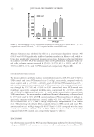
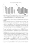
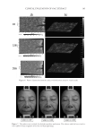
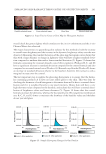
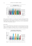
THE WHITENING AND ANTI-WRINKLE EFFECT OF PONEGRANATE 147 METHODS AND MATERIALS CHEMICALS A solution containing pomegranate was kindly provided by Health-love Therapeutics (Anyang, South Korea), who purchased concentrated pomegranate juice from Asya Fruit Juice and Food Ind. Inc. (Ankara, Turkey). The preparation of pomegranate solution was as follows: according to the manufacturer’s method, briefl y, the fruit was washed and then the rind was removed. Next, the pomegranate juice and seeds were separated using Bucher press. The pomegranate juice was sterilized in a high-temperature condition. The sterilized pomegranate juice was depectinized by pectinase. Next, it was fi ltered and concentrated which contains ellagic acid (2.31 mg/g). All test materials were maintained at 4°C until use. The solution was diluted with distilled water to concentrations of 0.005–0.5%. CELL CULTURES HaCaT cells were purchased from the American Type Culture Collection (ATCC, Manassas, VA) and cultured according to the manufacturer’s protocols. Cells were maintained in Dulbecco’s modifi ed Eagle’s medium (DMEM Sigma-Aldrich, St. Louis, MO) with 10% fetal bovine serum (FBS Lonza, Walkersville, MD), 100 μg/ml streptomycin (Sigma- Aldrich), and 100 U/ml penicillin (Sigma-Aldrich). HDF-N cell lines were obtained from ATCC and were cultured in fi broblast basal medium (FBM Lonza) supplemented with 2% FBS, 0.1% insulin, 0.1% recombinant human fi broblast growth factor (rhFGF), and 0.1% gentamicin. Melan-a cells were provided by Dr. Dorothy and were cultured in RPMI 1640 medium supplemented with 10% heat-inactivated FBS, 50 μg/ml strepto- mycin, and 50 U/ml penicillin. Cells were cultured at 37°C in a fully humidifi ed atmo- sphere of 5% CO2 and were passaged approximately every other day. WST-1 CELL PROLIFERATION ASSAY HaCaT, HDF-N, and Melan-a cells were plated at a density of 5 × 10⁴, 6 × 10³, and 9 × 10³ cells/well in a 96-well plate, respectively. The HaCaT, Melan-a, and HDF-N cells were exposed to various concentrations of PCS for 24 h and then cell proliferation reagent WST-1 (Roche, Mannheim, Germany) was added to each well and incubated in an incu- bator for 1 h. The absorbance of the wells at 450 nm was read using a microplate reader (TECAN, Männedorf, Switzerland). HYALURONAN ASSAY To determine the activity of hyaluronan synthesis, HaCaT cells were treated with 0.01%, 0.05%, and 0.1% PCS or N-acetyl-D-glucosamine (NAG) for 24 h, and then cells were trypsinized and counted for normalization. Hyaluronan concentration in the samples was quantifi ed using an enzyme-linked hyaluronan-binding protein sandwich assay (Cat no. DY3614 R&D Systems, Minneapolis, MN) based on the manufacturer’s methods (21).

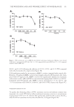
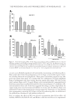
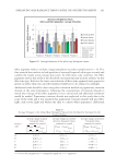
JOURNAL OF COSMETIC SCIENCE 148 ELASTASE INHIBITION ASSAY The elastase inhibition assay was performed by measuring the release of p-nitroaniline due to proteolysis of N-succinyl-(Ala)3-p-nitroanilide by human leucocyte elastase (Sigma-Aldrich) (22) in the presence or absence of PCS solution (0.05%, 0.1%, and 1%) or phosphoramidon (PPR, 10 μM) as a standard under exactly the same experimental conditions. The absorbance was measured at 405 nm with a 96-well microplate reader and the elastase inhibitory activity of each sample was calculated as follows: elastase in- hibitory activity (%) = 100-[(ODs/ODc) × 100], where ODs is the absorbance of the ex- perimental sample at 410 nm and ODc is the absorbance of the vehicle-treated control at 410 nm. The results are reported in terms of IC50 (the concentration at which the per- centage inhibition of elastase activity was 50%). MEASUREMENTS OF PROCOLLAGEN SYNTHESIS HDF-N cells were seeded in 48-well plates. Cells were treated with medium containing various concentrations of PCS solution (0.05%, 0.1%, and 1%) and then further culture for 24 hours. Culture medium was used for the determination of collagen synthesis. The collagen content was determined by procollagen type I C peptide enzyme-linked immu- nosorbent assay (ELISA) kit (MK101, Takara, Japan). TYROSINASE INHIBITION ASSAY Tyrosinase inhibition was tested according to Masamoto’s method (23). Briefl y, aliquots (0.05 ml) of PCS (0.05%, 0.1%, and 1%) were mixed with 0.5 ml of L -DOPA (Sigma- Aldrich) solution (1.25 mM) and 0.9 ml of sodium acetate buffer solution (0.05 M, pH 6.8), and preincubated at 25°C for 10 min. Then, 0.05 ml of an aqueous solution of mushroom tyrosinase (333 U/ml Sigma-Aldrich) was added to the mixture. This solu- tion was immediately monitored for the formation of dopachrome by measuring the lin- ear increase in optical density (OD) at 475 nm with a UV/V is spectrophotometer, and the tyrosinase inhibitory activity of each sample was calculated as follows: tyrosinase in- hibitory activity (%) = 100-[(ODs/ODc) × 100], where ODs is the absorbance of the ex- perimental sample at 475 nm and ODc is the absorbance of the vehicle-treated control at 475 nm. The results are reported in terms of IC50 (the concentration at which the percent- age inhibition of tyrosinase activity was 50%). Kojic acid (KA 1.25, 2.5, 5, 10, 20 and 40 μg/ml) was used as a standard under exactly the same experimental conditions. MELANIN FORMATION TEST IN MELAN-A CELLS Melanin content was measured according to the method of Hosoi (24) with slight modifi ca- tions. Melan-a cells were exposed to various concentrations of PCS solution (0.05%, 0.1%, and 1%). At the end of the treatment, the cells were lysed with 800 μl of 1 N NaOH (Merck KGaA, Darmstadt, Germany) containing 10% dimethyl sulfoxide (DMSO Sigma- Aldrich) for 1 h at 80°C. The absorbance at 400 nm was measured using a microplate
Purchased for the exclusive use of nofirst nolast (unknown)
From: SCC Media Library & Resource Center (library.scconline.org)