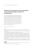
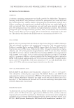
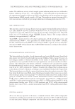
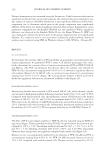
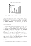
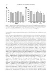
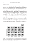
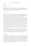
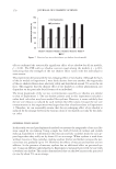
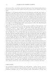
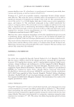
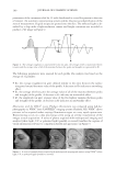
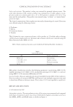
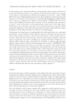
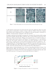
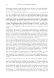
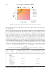
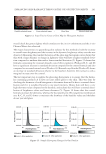
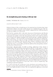
THE WHITENING AND ANTI-WRINKLE EFFECT OF PONEGRANATE 151 89.6%, and 6.3.6% following the addition of 0.01%, 0.05%, and 0.1% PCS compared with vehicle-treated cells, respectively (Figure 3B). UVA irradiation resulted in an increase in MMP-1 activity compared with control cells. However, UV-induced MMP-1 activity was strongly reduced in PCS-treated cells. Com- pared with UV-exposed cells, MMP-1 activity was signifi cantly inhibited on treatment with retinoic acid (1 μM) and PCS at concentrations ranging from 0.0001% to 0.001%. Compared with UV-exposed cells, MMP-1 activity increased by 137.1%, 69.3%, and 7.1% on treatment with 0.0001%, 0.0005%, and 0.001% PCS, respectively (Figure 3C). WHITENING EFFECTS OF PCS To explore the whitening effects of PCS, tyrosinase activity and melanin content were measured. As a result, PCS did not affect mushroom tyrosinase activity at concentrations ranging from 0.05% to 0.1%, whereas KA signifi cantly inhibited this activity. The IC50 value for KA on tyrosinase activity was calculated as 3.634 ppm (Figure 4A). Figure 1. PCS cytotoxicity. (A–C) HDF-N cells, HaCaT, and mouse melanocytes (Melan-a) were treated with various concentrations of PCS for 24 h. a p 0.01 compared with control (LSD test) b p 0.01 compared with the control (MW test).

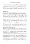
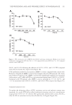
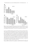
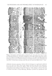
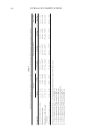
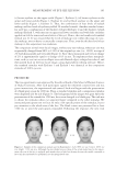
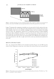
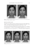
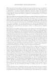
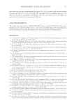
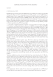
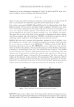
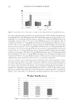
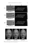
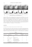
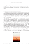
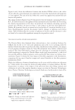
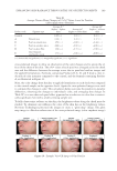
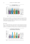
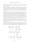
JOURNAL OF COSMETIC SCIENCE 152 Melanin formation was inhibited by PCS in a concentration-dependent manner. PCS (0.01% and 0.05%) signifi cantly inhibited melanin synthesis in Melan-a cells, while ar- butin also signifi cantly suppressed melanin production. Melanin production following the addition of 0.05% PCS was similar to that of 100 μM arbutin. Compared with con- trol cells, melanin content increased by 95.49 ± 9.27%, 86.3 ± 1.21%, and 67.12 ± 5.70% in 0.05%, 0.1%, and 1% PCS-treated cells, respectively (Figure 4B). HISTOMORPHOMETRIC CHANGES The mean number of epithelial surface microfolds decreased by 458.25% and −1.94% in UVB control and intact PCS-treated mice (1 ml/kg), respectively, compared with the intact control, and by −27.83%, −42.09%, and −35.83% in PCS-treated mice (0.5, 1, and 2 ml/kg, respectively) compared with UVB control mice. The mean epithelial thick- ness changed by 271.75% and −2.94% in UVB control and intact PCS-treated mice (1 ml/kg), respectively, compared with the intact control, and by −18.30%, −42.68%, and −32.62% in PCS-treated mice (0.5, 1, and 2 ml/kg, respectively) compared with UVB control mice. The mean number of infi ltrated dermal infl ammatory cells was altered by 1775.47% and −4.72% in UVB control and intact PCS-treated mice (1 ml/kg), re- spectively, compared with the intact control, and by −21.33%, −55.28%, and −49.09% in PCS-treated mice (0.5, 1, and 2 ml/kg, respectively) compared with UVB control mice. The percentage of collagen fi ber-occupied dermis in UVB control and intact PCS- administered mice (1 ml/kg) changed by 60.57% and −3.02% compared with the intact control, and −12.03%, −20.73%, and −16.58% in PCS-treated mice (0.5, 1, and 2 ml/kg) compared with UVB control mice, respectively (Figure 5, Table 1). DISCUSSION Our observations indicated that PCS increased hyaluronan synthesis but decreased elastase, collagenase, MMP-1, and tyrosinase activities, as well as melanin production. Thus, PCS Figure 2. Moisturizing effect of PCS. Hyaluronan synthesis was assessed in PCS-treated HaCaT. a p 0.01 compared with control (LSD test) b p 0.01 compared with the control (MW test).
Purchased for the exclusive use of nofirst nolast (unknown)
From: SCC Media Library & Resource Center (library.scconline.org)