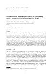
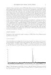
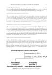
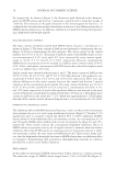
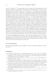
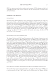
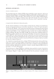
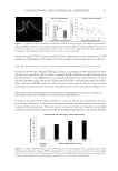
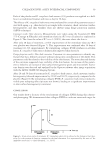
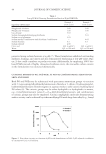
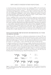
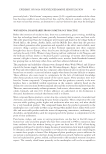
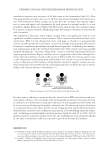
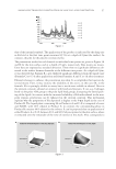
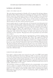
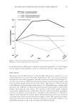
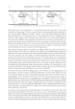
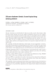
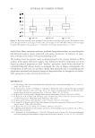
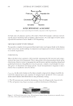
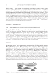
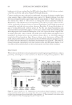
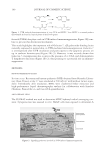
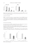
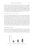
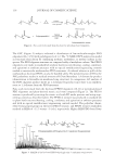
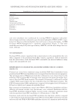
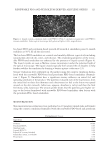
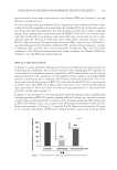
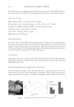
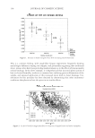
BATTLE AGAINST DANDRUFF 109 CFU/ml. On D0, a fi lter paper disk presoaked in 90 μl of the yeast mixture was applied (or not) for 24 h at the surface of skin explants, for a fi nal concentration of 1.1 × 10⁶ CFU/cm2. On D1, D2, and D4, explants were treated (or not) with a solution of 1.5% E. angustifo- lium extract (2μl/cm2). On D5, explants were collected. Observation of skin general mor- phology by optical microscopy was performed following staining of paraffi nized sections according to Masson’s trichrome Goldner variant. Immunostaining of hBD2 and hBD3 as well as TLR2 was done using antibodies specifi c for each proteins whose presence was subsequently revealed with secondary FITC-conjugate antibodies giving a green fl uores- cence. Cells nuclei were stained with propidium iodide. CLINICAL EVALUATION OF E. ANGUSTIFOLIUM EXTRACT A total of 24 volunteers (men and women, 20–61 years of age) with dandruff and greasy hair participated in the study. Following a 15 days wash-out period using a neutral shampoo, participants were instructed to wash their hair and scalp every 3 days for 30 days, with the same neutral shampoo to which was added 1.5% E. angustifolium extract. The pres- ence of dandruff was estimated on a scale of 0–5 by a trained technician, on D0, D3, D9, D15, and D30 (before shampooing). For this evaluation, the head was divided into four equal parts with a comb. The fi nal grade for dandruff presence on scalp was calculated as the mean of all four head part scores. Adherent and nonadherent dandruff were estimated separately. The presence of sebum on scalp was measured with the Sebumeter® SM 810 (Courage + Khazaka, Cologne, Germany) on D0, D15, and D30. Macrophotographs of scalp were additionally taken in standardized conditions on one preselected zone, at D0, D3, and D30. A self-assessment form was also fi lled out by all subjects on D30 to subjec- tively evaluate the properties of the study product (effi cacy, tolerance, and future use). RESULTS AND DISCUSSION Using a normal human sebocyte culture model and Nile red staining, the effect of E. angustifolium extract on lipid synthesis is presented in Figure 1, as percentages of relative fl uorescence units in comparison with DGAT (taken as 100%), a known inhibitor of lipid synthesis. As can be seen, E. angustifolium extract dose dependently inhibited sebum pro- duction. A substantial reduction by -43% was achieved with a 0.1% concentration of Figure 1. Epilobium angustifolium extract inhibits lipid synthesis in sebocytes.

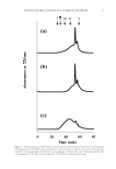
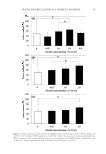
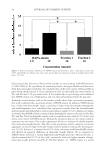
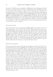
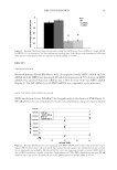
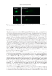
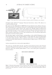
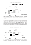
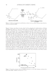
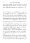
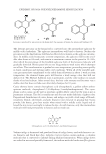
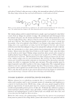
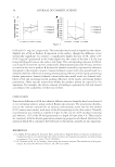
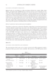
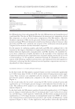
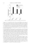
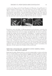
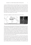
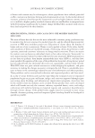
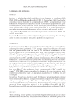
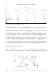
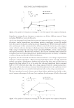
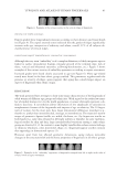
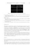
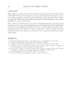
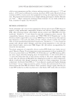
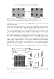
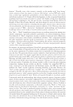
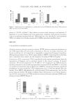
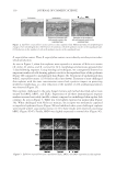
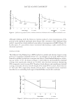
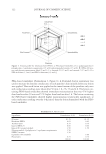
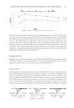
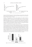
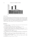
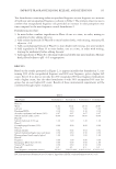
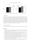
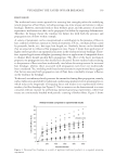
JOURNAL OF COSMETIC SCIENCE 110 E. angustifolium extract. Thus, E. angustifolium extract can act directly on sebocytes to reduce sebum production. As seen in Figure 2, when skin explants were exposed to a mixture of Malassezia strains (M. furfur, M. globosa, and M. restricta) for 24 h, morphological alterations appeared after 5 days following exposure. Using histological techniques, we consequently observed an important number of cells showing pyknotic nuclei in the suprabasal layer of the epidermis (Figure 2B) compared to unchallenged skin (Figure 2A). Treatment of unchallenged skin with E. angustifolium extract (1.5%) had no effect (not shown). Treatment of yeast-challenged skin explants with the same concentration extract had a positive impact on protecting epidermis morphology, as a clear reduction of the number of cells undergoing pyknosis was observed (Figure 2C). Skin explants challenged in the same fungal mixture and method described earlier were stained for hBD2, hBD3, and TLR2. Expressions of all three immunological response markers increased noticeably (middle column) compared to unchallenged skin explant (left column). As seen in Figure 3, TLR2 was very lightly expressed in normal skin (Figure 3A). When challenged with Malassezia mixture, the receptor was moderately expressed in superfi cial epidermal layers (Figure 3B) and inhibited when yeast-challenged explants were treated with E. angustifolium extract (1.5%). Same results were observed concerning hBD2 (Figure 3D–F). Finally, hBD3 was slightly expressed in normal skin (Figure 3G). Figure 2. Epilobium angustifolium extract protects skin explants from Malassezia-induced morphological changes. (A) Unchallenged skin. (B) Presence of numerous cells with pyknotic nuclei in the suprabasal layer. (C) Reduction in the number of cells with pyknotic nuclei in the suprabasal layer. Figure 3. Epilobium angustifolium extract modulates TLR2, hBD2, and hBD3 expression in skin explants.
Purchased for the exclusive use of nofirst nolast (unknown)
From: SCC Media Library & Resource Center (library.scconline.org)