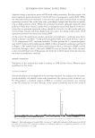
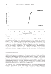
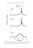
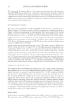
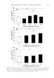
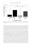
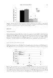
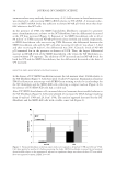
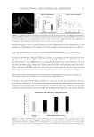
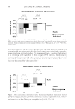
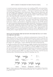
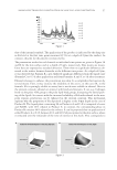
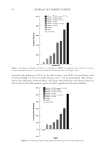
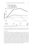
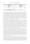
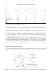
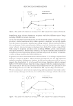
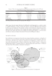
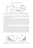
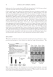
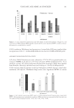
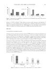
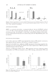
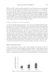
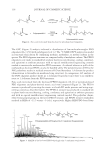
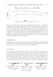
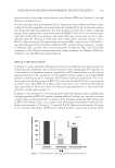
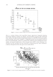
JOURNAL OF COSMETIC SCIENCE 22 such as a collagen (31). This gentle effect can be benefi cial in the development of useful biomaterials without any side effects. Surprisingly, a signifi cant difference in tyrosinase in- hibition was observed between fractions 1 and 2 (Figure 5). The average molecular weights of HAPA-elastin, fraction 1, and fraction 2, determined using gel fi ltration chromatogra- phy (Figure 3), were 4,700, 3,900, and 10,000 Da, respectively. The molecular weights were used to calculate the molarities of each sample. The molarities for a 10 mg/ml solution of HAPA-elastin, fraction 1, and fraction 2 were 2.1, 2.6, and 1.0 mM, respectively. Al- though the molarities of HAPA-elastin and fraction 1 at a concentration of 10 mg/ml were almost similar, HAPA-elastin showed relatively higher tyrosinase inhibition than fraction 1 did. Interestingly, fraction 2 showed relatively higher tyrosinase inhibition than HAPA- elastin and fraction 1 did, despite the low molarity of fraction 2 compared with either of them. It suggested that tyrosinase inhibition activity of elastin and elastin derivatives was not simply proportional to their molecular weight. It can be presumed that the observed difference in tyrosinase inhibition was due to the different sequences of amino acids in the various peptides and not merely due to the presence of different amino acids. It has been reported that no amino acid can, by itself, completely inhibit the activity of tyrosinase (32). Melanocytes have binding sites for the elastin-derived Val-Gly-Val-Ala-Pro-Gly (VGVAPG) hexapeptide. This hexapeptide is involved in bioactivities such as cell migration and prolif- eration (33). This may be the mechanism by which HAPA-elastin inhibited tyrosinase ac- tivity, which may subsequently promote skin cell turnover and result in whitening effects. In conclusion, HAPA-elastin is useful not only as a functional food but also as a cosmetic material, because it maintains the water content of the skin and inhibits tyrosinase activity. The fi ndings from this study may contribute to the development of better cosmetics with milder and more moisturizing effects on the skin. ACKNOWLEDGMENT We thank E&C HealthCare Ltd. and ECC Co., Ltd. for the fi nancial support to conduct this study. REFERENCES (1) C. M. Kielty, M. J. Sherratt, and C. A. Shuttleworth, Elastic fi bres, J. Cell Sci., 115, 2817–2828 (2002). (2) M. Hasegawa, T. Kawasaki, M. Saito, H. Ito, K. Okamoto, C. Arai, Y. Kashiwakura, and S. Yoshimura, Determination of aortic medial tissue elastin and collagen in stroke-prone spontaneously hypertensive rats, Nihon Ronen Igakkai Zasshi, 18(5), 336–341 (1981). (3) R. Yamaji, Development of Functional Materials for Diseases and Beauty in Women (CMC Publishing Co., Ltd. Tokyo, 2014), pp. 266–273. (4) T. Hayakawa, M. Sato, A. Saiga-Egusa, Y. Takahata, F. Morimatsu, and Y. Nomura, Effect of porcine arte- rial elastin peptide to the moisture content of mice skin, Nihon Chikusan Gakkaiho, 80(2), 215–222 (2009). (5) S. G. Harvey, J. R. Gibson, and C. A. Burke, L-cysteine, glycine and dl-threonine in the treatment of hypostatic leg ulceration: A placebo-controlled study, Pharmatherapeutica, 4(4), 227–230 (1985). (6) H. Ohara, S. Ichikawa, H. Matsumoto, M. Akiyama, N. Fujimoto, T. Kobayashi, and S. Tajima, Collagen- derived dipeptide, proline-hydroxyproline, stimulates cell proliferation and hyaluronic acid synthesis in cultured human dermal fi broblasts, J. Dermatol., 37(4), 330–338 (2010). (7) H. W. Spier and G. Pascher, Analytical and functional physiology of the skin surface, Hautarzt, 7(2), 55–60 (1956). (8) H. Sage and W. R. Gray, Studies on the evolution of elastin—I. Phylogenetic distribution, Comp. Bio- chem. Physiol. B, 64(4), 313–327 (1979).

WATER-SOLUBLE ELASTIN AS A COSMETIC MATERIAL 23 (9) R. P. Mecham and G. Lange, Antigenicity of elastin: Characterization of major antigenic determinants on purifi ed insoluble elastin, Biochemistry, 21(4), 669–673 (1982). (10) M. Katoh, F. Hamajima, T. Ogasawara, and K. Hata, Assessment of human epidermal model LabCyte EPI-MODEL for in vitro skin irritation testing according to European Centre for the Validation of Alternative Methods (ECVAM)-validated protocol, J. Toxicol. Sci., 34(3), 327–334 (2009). (11) M. Katoh, F. Hamajima, T. Ogasawara, and K. Hata, Assessment of the human epidermal model Lab- Cyte EPI-MODEL for In vitro skin corrosion testing according to the OECD test guideline 431, J. Toxicol. Sci., 35(3), 411–417 (2010). (12) T. Hikima, N. Kaneda, K. Matsuo, and K. Tojo, Prediction of percutaneous absorption in human using three- dimensional human cultured epidermis LabCyte EPI-MODEL, Biol. Pharm. Bull., 35(3), 362–368 (2012). (13) Y. Tokudome, M. Katayanagi, and F. Hashimoto, Esterase activity and intracellular localization in re- constructed human epidermal cultured skin models, Ann. Dermatol., 27(3), 269–274 (2015). (14) A. Adhikari, H. P. Devkota, A. Takano, K. Masuda, T. Nakane, and P. Basnet, Screening of Nepalese crude drugs traditionally used to treat hyperpigmentation: In vitro tyrosinase inhibition, Int. J. Cosmet. Sci., 30(5), 353–360 (2008). (15) E. Shiratsuchi, M. Ura, M. Nakaba, I. Maeda, and K. Okamoto, Elastin peptides prepared from piscine and mammalian elastic tissues inhibit collagen-induced platelet aggregation and stimulate migration and proliferation of human skin fi broblasts, J. Pept. Sci., 16(11), 652–658 (2010). (16) L. B. Sandberg, Elastin structure in health and disease, Int. Rev. Connect Tissue Res., 7, 159–210 (1976). (17) R. C. Burke, T. H. Lee, and V. Buettner-Janusch, Free amino acids and water soluble peptides in stratum corneum and skin surface fi lm in human beings, Yale J. Biol. Med., 38(4), 355–373 (1966). (18) Japan Health and Nutrition Food Association, A Guide to the Dietary Food Supplement ELASTIN, accessed September 9, 2013, http://www.jhnfa.org/ (19) R. S. Rapaka and D. W. Urry, Coacervation of sequential polypeptide models of tropoelastin. Synthesis of H-(Val-Ala-Pro-Gly)n-Val-OMe and H-(Val-Pro-Gly-Gly)n-Val-OMe, Int. J. Pept. Protein Res., 11(2), 97–108 (1978). (20) D. W. Urry, Protein elasticity based on conformations of sequential polypeptides: The biological elastic fi ber, J. Protein Chem., 3(5), 403–436 (1984). (21) K. Kaibara, K. Miyakawa, K. Okamoto, Y. Uemura, and M. Kondo, Development of biomaterials cor- responding to coacervate characteristics, Kobunshi Ronbunshu, 48(5), 311–317 (1991). (22) D. W. Urry, T. L. Trapane, R. B. McMichens, M. Iqbal, R. D. Harris, and K. U. Prasad, Nitrogen-15 NMR relaxation study of inverse temperature transitions in elastin polypentapeptide and its cross- linked elastomer, Biopolymers, 25, S209–228 (1986). (23) J. D. Bos and M. M. Meinardi, The 500 Dalton rule for the skin penetration of chemical compounds and drugs, Exp. Dermatol., 9(3), 165–169 (2000). (24) T. J. Brown, D. Alcorn, and J. R. Fraser, Absorption of hyaluronan applied to the surface of intact skin, J. Invest. Dermatol., 113(5), 740–746 (1999). (25) C. Kawada, T. Yoshida, H. Yoshida, W. Sakamoto, W. Odanaka, T. Sato, T. Yamasaki, T. Kanemitsu, Y. Masuda, and O. Urushibata, Ingestion of hyaluronans (molecular weights 800 k and 300 k) improves dry skin conditions: a randomized, double blind, controlled study, J. Clin. Biochem. Nutr., 56(1), 66–73 (2015). (26) S. Briganti, E. Camera, and M. Picardo, Chemical and instrumental approaches to treat hyperpigmenta- tion, Pigment Cell Res., 16(2), 101–110 (2003). (27) P. A. Riley, Melanin, Int. J. Biochem. Cell Biol., 29(11), 1235–1239 (1997). (28) T. S. Chang, An updated review of tyrosinase inhibitors, Int. J. Mol. Sci., 10(6), 2440–2475 (2009). (29) M. Schurink, W. J. van Berkel, H. J. Wichers, and C. G. Boeriu, Novel peptides with tyrosinase inhibitory activity, Peptides, 28(3), 485–495 (2007). (30) N. W. Hsiao, T. S. Tseng, Y. C. Lee, W. C. Chen, H. H. Lin, Y. R. Chen, Y. T. Wang, H. J. Hsu, and K. C. Tsai, Serendipitous discovery of short peptides from natural products as tyrosinase inhibitors, J. Chem. Inf. Model., 54(11), 3099–3111 (2014). (31) Y. L. Zhuang, X. Zhao, and B. F. Li, Optimization of antioxidant activity by response surface methodol- ogy in hydrolysates of jellyfi sh (Rhopilema esculentum) umbrella collagen, J. Zhejiang Univ. Sci. B., 10(8), 572–579 (2009). (32) M. Ishikawa, I. Kawase, and F. Ishii, Combination of amino acids reduces pigmentation in B16F0 melanoma cells, Biol. Pharm. Bull., 30(4), 677–681 (2007). (33) C. H. Chang, Y. Kawa, R. K. Tsai, J. H. Shieh, J. W. Lee, H. Watabe, T. Kawakami, Y. Soma, S. Tajima, and M. Mizoguchi, Melanocyte precursors express elastin binding protein and elastin-derived peptide (VGVAPG) stimulates their melanogenesis and dendrite formation, J. Dermatol. Sci., 51(3), 158–170 (2008).
Purchased for the exclusive use of nofirst nolast (unknown)
From: SCC Media Library & Resource Center (library.scconline.org)