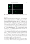
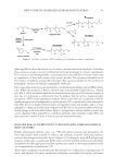
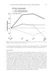
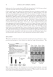
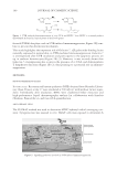
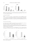
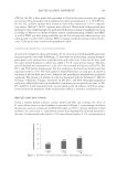
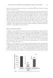
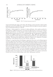
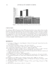
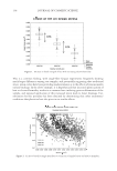
JOURNAL OF COSMETIC SCIENCE 26 to have strong antiaging benefi ts as SIRT6-defi cient mice develop a progeroid-like syn- drome resembling premature aging and die after 4 weeks (6). Mammalian cells defi cient in SIRT6 have defects in DNA repair resulting in genomic instability. These include base excision repair (BER) (6,7,8), double strand break (DSB) repair (7,9–12), and telomere maintenance (7,13,14). Current evidence indicates that SIRT6 regulates BER either by modulating BER factors or by regulating the chromatin density permitting the accessibility of BER factors to DNA damage sites (7,13). Success- ful DSB repair requires that SIRT6 recruit a chromatin remodeler, SNF2H, to the DSB (11) and the deacetylation of a C-terminal binding protein, CtIP, to promote DNA- end resectioning (10). Proper telomere maintenance requires the binding of SIRT6 to telomeric chromatin resulting in the deacetylation of H3K9 histones and preventing chromosome end fusion. Thus, SIRT6 functions both as an enzyme and as a scaffold for establishing a unique microenvironment for recruiting other proteins needed for DNA repair. In addition to DNA repair, SIRT6 is involved in infl ammation control (3). Pyrimidine dimers are the primary trigger of post-ultraviolet (UV) erythema and their removal requires DNA nicking in order to mediate excision repair (15). SIRT6 promotes tumor necrosis factor (TNF)-α secretion by deacylating two myristoyl groups from lysine residues at peptide positions 19 and 20 of TNF-α (16). Conversely, SIRT6 is recruited to promoter regions of nuclear factor (NF)-κB target genes and physically docks with the NF-κB subunit, RelA (5,17). SIRT6 silences these NF-κB target genes by modifying the chromatin via deacetylation of H3K9 on nearby histones (5,17). The altered chromatin structure destabilizes RelA binding to the target gene promoter, terminating NF-κB stimulation of the target genes. A SIRT6-defi cient mouse survives only 30 days because both NF-κB genes are unregulated leading to hyperactive NF-kB signaling (18). A SIRT6-defi cient mouse crossed with a RelA +/− mouse rescues early lethality of SIRT6-defi cient mice, giving rise to progeny which survive for more than 100 days, indicating that a reduction of the NF-κB copy number results in longer survival (17,18). Further, recall that transgenic male mice overexpressing SIRT6 have increased longevity, about 15% (19), which is associated with the insulin-like growth factor 1 signaling pathway (3). As mentioned earlier, a reduced capacity to maintain genomic integrity and an increased sensitivity to oxidative stress and infl ammation are causes of premature aging in the SIRT6 knockout mouse. SIRT6-defi cient mice, as well as several mouse models of nucle- otide excision repair defi ciency, exhibit a premature aging phenotype, which is associated with insulin signaling (16). This premature aging phenotype in mice is associated with dysregulation of insulin signaling as a result of glucose metabolism dysfunction (16,20), hyperactive NF-κB signaling (17), and genomic instability (7). SIRT6 is involved in all these pathways. With regard to humans, mesenchymal stem cells defi cient in SIRT6 have dysregulated redox metabolism because SIRT6 functions as a transactivator for nuclear factor erythroid 2-related factor 2, establishing a connection between SIRT6 and oxidative stress (21). The imbalanced relationship between DNA repair and metabolism in SIRT6- defi cient mice is an important theme in premature aging. The upregulation of NF-κB as a consequence of SIRT6 defi ciency further illustrates the multiple connections of SIRT6 with premature aging. We sought to investigate the importance of DNA repair and NF-κB in a tissue culture model. We explored the effects of SIRT6 knockdown in human dermal fi broblasts under normal conditions and after environmental stress from ultraviolet B (UVB) and ultraviolet A (UVA) irradiation. In this report, we evaluated the effectiveness of

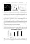
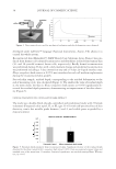
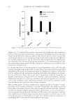
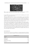
SIRT6 KNOCKDOWN 27 SIRT6 as a protective molecule in relation to the extent of DNA damage and the level of NF-κB expressed under normal conditions and after UV exposure of normal human dermal fi broblasts. MATERIALS AND METHODS TISSUE CULTURE Neonatal human dermal fi broblasts were purchased from Life Technologies. Cells were maintained in Dulbecco Modifi ed Eagle's Media (DMEM Life Technologies, Grand Island, NY) and 10% fetal bovine serum (FBS) (HyClone, GE Heathcare, Logan, UT). Cells were grown at 37ºC and 5% humidity. Donor 828840 was used at p5 and at p8 for the SIRT6 electroporation, comparing SIRT6 messenger RNA (mRNA) levels relative to RPLPO mRNA levels. Donor 871299 was used for the NF-κB experiment at passage 5. Donor 904886 was used for the fi rst comet assay, 40 mJ/cm2 UVB and 5, 10, or 20 J/m2 UVA, at passage 12. Donor 871299 was used for the second comet assay, 40 mJ/cm2 UVB and 20 J/m2 UVA, at passage 6. ELECTROPORATION OF SMALL-INTERFERING RNA The Amaxa Nucleofector II/2b Lonza Electroporation Kit (Cologne, Germany) was used to deliver small-interfering RNA (siRNA) specifi c for SIRT6. Cells were washed with Hank’s buffered saline solution before trypsinizing. An optimized electroporation pro- tocol, ID 83, from Lonza was followed. The fi broblast pellet was resuspended in the appropriate buffer and supplement (Lonza electroporation kit, VPD-1001). The Nucleo- fector II/2b was set to program U-020. Silencing was achieved using an ON-TARGETplus SMARTpool for SIRT6 from Dharmacon (Lafayette, CO) containing the following se- quences: 5′-CCAAGUGUAAGACGCAGUA-3′, 5′-GUACAUCGCUGCAGAUCCG-3′, 5′-CCAAAAGGGUGAAGGCCAA-3′, and 5′-GAACUGGCGAGGCUGGUCU-5′ or the single sequence synthesized from Dharmacon, 5′-GGAACAUGUUUGUGGAAGAUU-3′ (22). The ON-TARGETplus nontargeting (NT) pool from Dharmacon included the four following sequences: 5′-UGGUUUACAUGUCGACUAA-3′, 5′-UGGUUU ACAUGUUGUGUGA-3′, 5′-UGGUUUACAUGUUUUCUGA-3′, and 5′-UGGUUUA CAUGUUUUCCUA-3′. Each electroporation included 8–10 × 105 fi broblasts. Prior to electroporation, each cell pellet was resuspended by adding a volume of 105 μl of Lonza buffer containing siRNA at a fi nal concentration of 1 μM. The resuspended pellet was transferred to a cuvette for electroporation. Electroporated fi broblasts were seeded in four wells of a six-well plate or a 100-mm plate and incubated in 10% FBS and DMEM. RNA PURIFICATION AND REAL-TIME REVERSE TRANSCRIPTION POLYMERASE CHAIN REACTION Neonatal fi broblasts were washed with Dulbecco’s phosphate-buffered saline (DPBS) prior to extracting with 600 μl of RLT buffer from the RNeasy mini kit (Qiagen, Hilden,
Purchased for the exclusive use of nofirst nolast (unknown)
From: SCC Media Library & Resource Center (library.scconline.org)