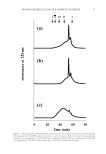
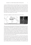
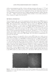
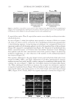
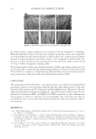
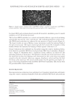
JOURNAL OF COSMETIC SCIENCE 28 Germany). The RNA extract was applied to a QIAshredder column (Qiagen) and eluted. The extract was further purifi ed according to the RNeasy protocol. The RNA concentra- tion was determined by absorbance at 260 nm with a Beckmann DU-7000 (Brea, CA) spectrophotometer (1A = 40 μg/ml). RNA was reverse transcribed with a high-capacity complementary DNA reverse transcription kit (Applied Biosystems, Foster City, CA) and amplifi ed with Taqman primer/probe sets from Applied Biosystems specifi c for human SIRT6, assay ID Hs00966001_m1, and RPLPO (60S acidic ribosomal protein P0), assay ID Hs00420895_gH. Real-time reverse transcription polymerase chain reaction (PCR) was performed and analyzed on an Applied Biosystems 7500 Fast instrument. Each PCR cycle involved 3 s at 95°C for melting and 60 s at 60°C for annealing and extension. A total of 40 cycles were completed. MODULATION OF NF-ΚB Neonatal fi broblasts were electroporated with SIRT6 siRNA and incubated for 48 h (21) before exposing the cells to 40 or 80 mJ/cm2 UVB. These doses were not cytotoxic to neonatal fi broblasts (data not shown). An irradiation chamber, BS-03 (manufactured by Dr. Grőbel UV-Elektronik GmbH, Ettlingen, Germany), was used to deliver the UVB. Six hours after irradiation, the cells were lysed and the total protein was extracted with NE-PER nuclear and cytoplasmic extraction kit from Thermo Fisher Scientifi c (Waltham, MA). Total protein was quantitated with a Micro BCA protein assay kit from Thermo Fisher Scientifi c. A chemiluminescent transcription factor detection kit from Thermo Fisher Scientifi c was used to detect NF-κB p50. A Molecular Devices Spectra- Max M2e (Sunnyvale, CA) was used to measure chemiluminescence. DETECTION OF DNA DAMAGE Each well of a Trevigen comet slide (Gaithersburg, MD) was seeded with 10,000 electro- porated fi broblasts in 200 μl of DMEM and 10% FBS. The neonatal fi broblasts were previously electroporated with SIRT6 siRNA or NT siRNA (NT). After 48 h of growth, neonatal fi broblasts were exposed to 40 mJ/cm2 UVB and 20 J/cm2 UVA using the BS-03 irradiation chamber. Cells were exposed to UV at room temperature. Cells were grown for another 4 h at 37°C and 5% humidity before processing the slides with Trevigen re- agents. No inhibitors of DNA repair were added. Slides were washed with DPBS and 75 μl of melted agarose was pipetted on each well. The cells were then processed accord- ing to the Trevigen protocol with the following changes. After 10 min at 4°C, cells were lysed for 3 h on ice. Slides were removed from the lysis solution and placed into the alka- line solution for 30 min. Slides were electrophoresed in a cold alkaline solution (300 mM NaOH, 1 mM ethylenediaminetetraacetic acid, pH 13) for 30 min at 23 V. After elec- trophoresis, slides were rinsed with water, fi xed with 70% ethanol for 5 min, and dried overnight. DNA was detected with 50 μl SYBR Green (Thermo Fisher) per well (diluted 1:10,000 in TE buffer) and incubated for 5 min at 4°C. Slides were viewed under an Olympus BX51 microscope (Center Valley, PA) using a Fluorescein Isothiocyanate fi lter and a 20× objective. Images were captured using Nikon Elements Software (Melville, NY). The tail moments were determined with Comet Score software from TriTek (Wilmington, DE).

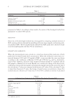
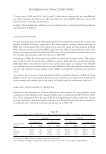
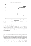
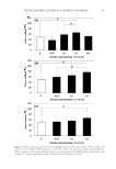
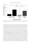
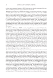
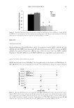
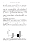
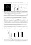
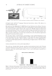
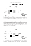
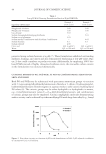
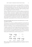
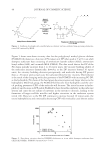
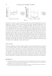
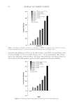
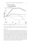
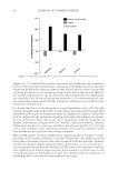
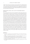
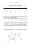
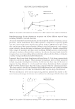
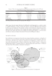
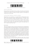
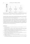
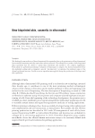
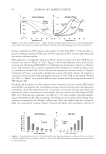
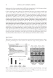
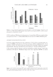
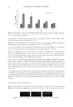
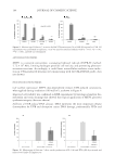
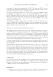
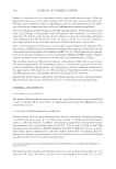
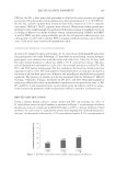
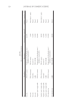
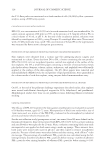
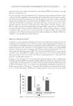
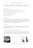
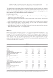
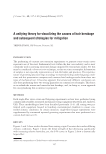
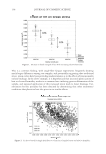
SIRT6 KNOCKDOWN 29 RESULTS SIRT6 KNOCKDOWN Neonatal human dermal fi broblasts were electroporated with SIRT6 siRNA and the mRNA levels for SIRT6 were monitored 48 h after electroporation. A 72% decrease in SIRT6 mRNA was achieved with four siRNAs and a 68% decrease with one SIRT6 siRNA (Figure 1). The NT siRNA level of SIRT6 mRNA was comparable to the untreated. IMPACT OF SIRT6 KNOCKDOWN ON NF-κB SIRT6 knockdown elevates NF-κB p50 levels signifi cantly in the absence of UVB (Figure 2). NF-κB p50 levels were monitored 52 h after electroporation using an enzyme-linked Figure 1. Neonatal fi broblasts were electroporated using four siRNAs specifi c for SIRT6 or a single siRNA for SIRT6 (22). An average of 70% knockdown was observed after 48 h relative to the nontargeted cells from three separate experiments. Data are expressed as mean ± SEM, *p 0.005. Figure 2. Neonatal fi broblasts were electroporated with SIRT6 siRNA and incubated for 48 h before expos- ing to UVB. Six hours later, cells were lysed. The nuclear fraction (squares) was separated from the cytosolic faction (diamonds) and probed for NF-κB p50 (NT-cy, -nu = non-targeting cytosolic or nuclear SIRT6 KD-cy, -nu = SIRT6 knockdown cytosolic or nuclear). In the absence of UVB, SIRT6 knockdown increased NF-κB translocation to the nucleus fourfold relative to NT. Data expressed as mean ± SEM, *p 0.05.
Purchased for the exclusive use of nofirst nolast (unknown)
From: SCC Media Library & Resource Center (library.scconline.org)