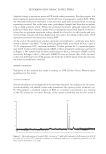
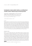
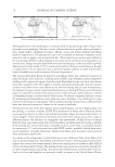
J. Cosmet. Sci., 68, 35–41 ( January/February 2017) 35 Collagen XVIII: A key interfacial component of the skin architecture ISABELLE BONNET, LARA JOBEILI, SÉBATIEN CADAU., NICOLAS BERTHÉLÉMY., AUDREY PIERROT, CARINE TEDESCHI, VINCENT BARDEY, GUILLAUME FARGIER, DELPHINE RIVAL, CHRISTINE JEANMAIRE, CATHERINE BONNAUD-ROSAYE, WENDY CHAN, MANASI CHAVAN, VALÉRIE ANDRÉ-FREI, HASSAN ZAHOUANI, and PATRICIA ROUSSELLE, BASF Beauty Care Solutions France, Lyon, France (B.I., C.S., B.N., P.A., T.C., B.V., F.G., R.D., J.C., B.C., A.V.), BASF Corporation, Tarrytown, NY 10591 (C.W., C.M.), Tribology and System Dynamics Laboratory’s, Ecole Centrale Lyon, Ecully, France (Z.H.), and Laboratoire de Biologie Tissulaire et Ingénierie Thérapeutique, CNRS, Université Lyon 1, Lyon, France (L.J., R.P.). INTRODUCTION Collagen XVIII belongs to the multiplexins, known as extracellular matrix proteins that contain multiple triple-helix domains (collagenous domains) interrupted by noncol- lagenous domains. Besides, as collagen XVIII is a basement membrane (BM) heparan sulfate proteoglycan, it holds the structural properties of both collagen and proteoglycan. This collagen is expressed ubiquitously in various BM structures throughout the body. In skin and compared to other collagen types, collagen XVIII displays the broadest repar- tition as it could be synthetized by keratinocytes, endothelial cells, epithelial cells of the sweat glands and hair follicles stem cells, and adipocytes (1). However, the complete physiological role of collagen XVIII is not fully understood, even if its localization and ultrastructural organization reveal that it is an important component of all BM molecular networks present in skin (2). Understanding its expression modulation with age is of great interest for cosmetic research and could provide new strategies to counteract the loss of tissue structure and cohesion seen during aging of epidermis, dermis, hypodermis, and scalp. Address all correspondence to Valerie Andre-Frei at valerie.andre-frei@basf.com.

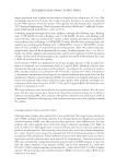
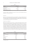
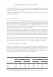
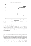
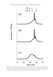
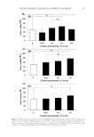
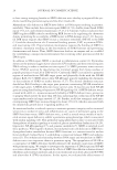
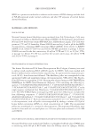
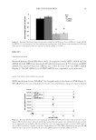
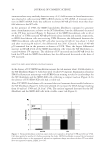
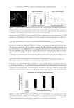
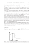
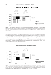
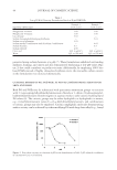
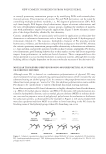
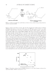
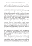
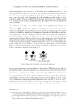
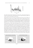
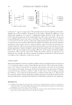
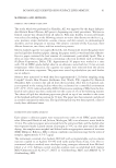
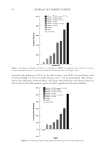
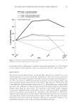
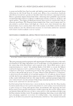
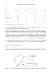
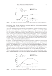
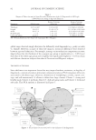
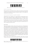
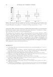
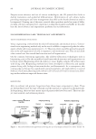
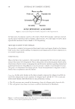
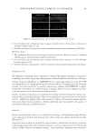
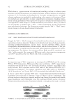
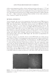
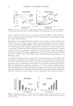
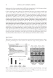
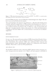
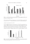
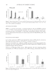
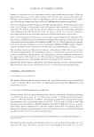
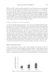
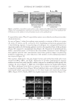
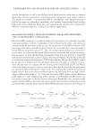
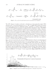
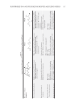
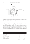
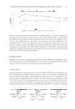
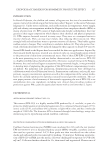
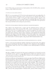
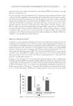
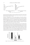
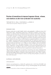
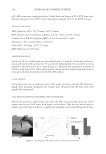
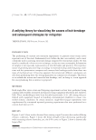
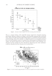
JOURNAL OF COSMETIC SCIENCE 36 METHODS AND RESULTS SPECIFIC ANTIBODY DESIGN To specifi cally target the collagen XVIII portion involved in tissue structure, and not its endostatin domain (Figure 1), we designed an affi nity-purifi ed rabbit antibody against its two collagenic domains: COL1 and COL 5. This antibody was used for collagen XVIII immuno- localization on skin biopsies of different ages and for the design of an enzyme-linked immunosorbent assay (ELISA) on keratinocytes allowing cosmetic ingredient screening. COLLAGEN XVIII EXPRESSION DURING SKIN AGING At fi rst immuno-labelling of 12 skin samples coming from face lifting was performed to localize and quantify this protein in the skin structure (n = 6 for 8–15 years old and for 30–69 years old). Briefl y, 7-μm cryosections were fi xed using cold methanol/acetone during 10 min and unspecifi c sites were blocked using 10% normal goat serum diluted in phosphate- buffered saline (PBS) at room temperature. Then sections were incubated within the primary antibody and after two washes in PBS, the sections were incubated with Cy3- labelled anti-rabbit antibody and counterstained with Dapi. Sections were examined using a Zeiss LSM 700 confocal microscope and the acquisition was realized using the Z-stack mode of the Zen program. Quantifi cation data are presented as mean values and standard error of the mean from at least three measures performed on biological triplicate samples for each skin samples (Figure 2). As compared to young skin specimens, collagen XVIII staining was less intense and homog- enous. In a 60 years old skin sample, heterogeneity of the lamina densa labeling was observed as well as zones where the lamina densa starts to duplicate and detach from the dermal epidermal. Quantifi cation allowed to evidence a strong collagen XVIII decrease as skin ages (-43.5% for older group). The age decrease of collagen XVIII was confi rmed using quantitative reverse transcription polymerase chain reaction (qRT-PCR) study on keratinocytes extracted from 50 abdominal skin biopsies (Figure 2, right), which evidenced that the proteomic decrease occurs Figure 1. Collagen XVIII structure (NC: non collagenic domains, COL: collagenic domains, : antigenic area).
Purchased for the exclusive use of nofirst nolast (unknown)
From: SCC Media Library & Resource Center (library.scconline.org)