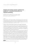
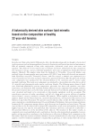
J. Cosmet. Sci., 68, 85–90 ( January/February 2017) 85 New bioprinted skin, cosmetic in vitro model SEBASTIEN CADAU, DELPHINE RIVAL, VALERIE ANDRE-FREI, MANASI CHAVAN M, DELPHINE FAYOL, MARINE SALDUCCI, BRUNO BRISSON, and FABIEN GUILLEMOT, BASF Beauty Care Solutions, Lyon, France (S.C., D.R., V.A.), Poietis, Pessac, France (D.F., B.B., F.G.), and BASF Corporation, Tarrytown, NY 10591 (M.C.). Synopsis We developed a new evolution of three-dimensional skin equivalent due to the optimization of four-dimensional laser-assisted bioprinting and skin equivalent culture protocols. This allowed us to produce fully bioprinted skin equivalents that are closed to current skin equivalents and suitable to test cosmetic ingredients. Particularly, we performed preliminary evaluation of maturogens to improve the dermis maturation before the epidermal seeding and we designed a specifi c “micropattern” to reproduce the nonlinear aspect of the dermal–epidermal junction. Finally an active ingredient was applied during the production of the bioprinted skin equivalent. INTRODUCTION Although three-dimensional (3D) printing itself is a relatively new technology, invented three decades ago, it contributes to one of the most promising medical technological advance of the century in bioscience and its market potential is only just beginning to be realized in the case of bioprinting. The fi rst description of bioprinting occurred in 1988 when R. J. Klebe described Cytoscribing, the fi rst two and 3D synthetic tissues construction on fi bronectin substrate using ink-jet printer and computer-assisted high-precision posi- tioning of cells. From the beginning of the technology development, the promises are to establish in a short timeframe precise spatial arrangements within large populations of cells to resemble natural tissues and organs for regenerative medicine or testing applications. Different printing technologies have been successfully developed using either sophisticated and complementary ink-jet, bioextrusion, or laser printers. For example, the fi rst 3D skin printing production was described in 2012 with 3D arrangement of vital cells by laser- assisted bioprinting (LaBP) as multicellular fi broblasts and keratinocytes embedded in collagen for in vitro testing application. In 2013, PrintAlive Bioprinter using complex microfl uidic device has allowed human microtissue arrays to be routinely defi ned with unprecedented speed and resolution for grafting application. Address all correspondence to Valerie Andre-Frei at valerie.andre-frei@basf.com.

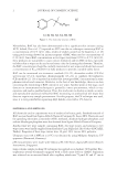
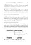
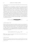
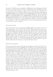
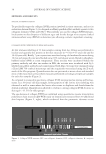
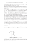
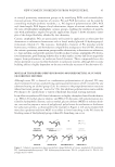
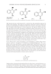
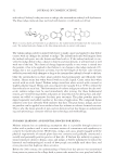
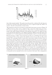
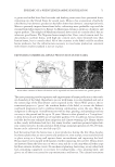
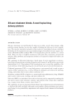
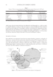
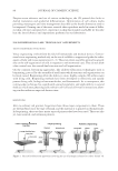
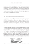
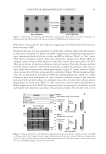
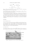
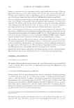
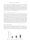
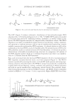
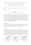
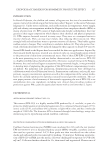
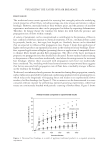
JOURNAL OF COSMETIC SCIENCE 86 Despite recent advances and use of various technologies, the 3D printed skin lacks in dermal maturation and epidermal differentiation. Optimization of cell culture media providing maturogens and time management described as the fourth dimension need to be improved. Owning one of the most versatile skin equivalent model in terms of differ- ent skin cell types integrated yet, experience in skin functionality and LAB, we describe here the latest advances and experiments performed in our laboratories. FOUR-DIMENSIONAL LaBP: TECHNOLOGY AND BENEFITS TISSUE ENGINEERING EVOLUTIONS Tissue engineering evolved from the fi eld of biomaterials and medical devices. Conven- tional tissue engineering methods rely on the use of scaffolds to support and guide the subse- quent cellular and tissue organization (1–3). These top-down assembly approaches greatly rely on the self-organization of cells in response to environmental cues. They do not allow a fi ne control over the created fi nal structure and cell organization. On the contrary, bottom-up approaches, like additive fabrication technologies such as bioprinting, proceed by the assembly of small units which structure and organization can be fi nely tuned. Bioprinting offers the ability to create highly complex 3D architectures with living cells. Bioprinting methods have been developed to effectively and rapidly pattern living cells, biological macromolecules, and biomaterials. As a consequence, this cutting-edge technique has signifi cantly gained popularity and applicability in several fi elds as it facilitates physiologically relevant cell–cell and cell–matrix interactions allow- ing studies within an expected shorten time. BIOPRINTING Akin to ordinary ink printers, bioprinters have three major components to them. These are the hardware used, the type of bioink, and the material it is printed on (biomaterials). In bioprinting, there are three major types of printers that have been used. These are ink- jet, laser-assisted, and extrusion printers. Figure 1. Selected biofabrication approaches involving the use of hydrogels in form of so-called “bioink” (4).
Purchased for the exclusive use of nofirst nolast (unknown)
From: SCC Media Library & Resource Center (library.scconline.org)