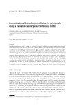
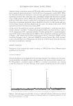
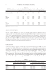
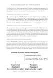
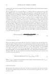
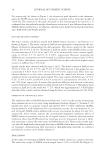
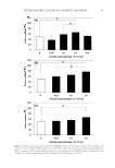
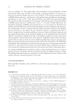
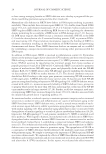
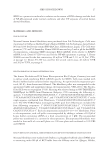
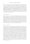
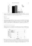
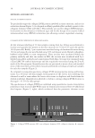
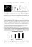
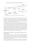
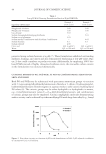
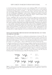
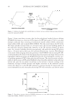
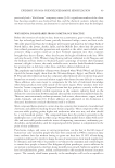
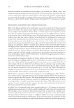
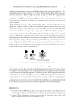
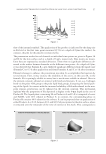
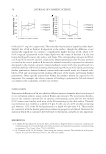
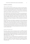
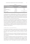
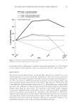
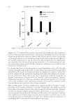
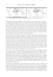
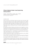
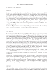
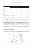
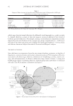
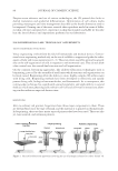
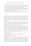
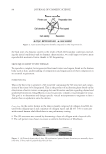
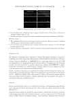
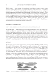
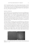
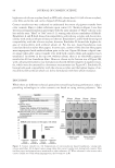
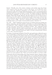
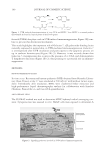
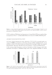
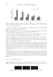
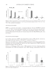
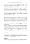
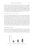
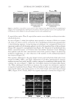
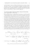
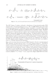
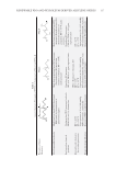
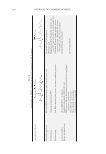
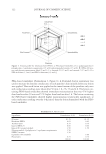
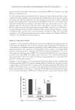
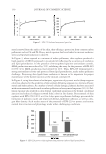
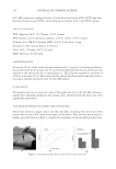
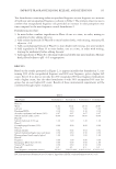
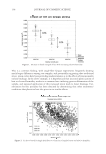
NEW BIOPRINTED SKIN, COSMETIC IN VITRO MODEL 87 ▪ Laser-assisted bioprinters are less common and use lasers focused on an absorbing substrate to generate pressures that propel cell-containing materials onto a collector substrate. Printers that utilize lasers provide high-resolution printing and because it is a nozzle-free device, clogging of the nozzle is avoided (Figure 1, left). ▪ Thermal ink-jet printers electrically heat the printhead to produce air-pressure pulses that force droplets from the nozzle, whereas acoustic printers use pulses formed by piezoelectric or ultrasound pressure (Figure 1, middle). Ink-jet printers are mainly used in bioprinting for fast and large-scale products. ▪ Microextrusion printers use pneumatic or mechanical (piston or screw) dispensing systems to extrude continuous beads of material and/or cells (Figure 1, right). LABP TECHNOLOGY 3D laser-assisted bioprinter has a near infrared pulse laser source and a focus system to adjust the ejecta size. A laser is beamed through a transparent slide coated with an absorbent layer, enabling light energy to be converted into kinetic energy. A thin matrix layer, con- taining the component to be printed and a recipient substrate, is positioned a few microns away from the fi rst slide. Laser pulses are programmed to be sent approximately every nanosecond. This generates inkjets (cell containing mini-droplets), which are deposited layer by layer. In this system, physical ejection conditions—energy and viscosity—as well as droplet volume to around picoliter accuracy are controlled. The biological ink cartridge scans quickly, generating over 10,000 droplets a second with a resolution of 20 μm. Compared to man- ual skin equivalent production, the time to make a biological structure 1 cm2 and 200– 300 μm thick useful for in vitro testing is reduced by two-thirds. Our preliminary studies have shown that printable extracellular matrix and cells can be combined in a laser-assisted printing sequence to fabricate a stable and organized soft free form tissue, which can host a high cell density de novo. The LaBP can print versatile bio- logical patterns such as cell clusters, cell confl uent surface, and cell alignments according to computer-aided design. Also, a cell-level resolution of cell printing at a high speed (5 kHz) is achievable by this laser-assisted bioprinter. Such precision and speed were a prerequisite to apply the LaBP to cellularized tissue fabrication (5). As a matter of facts, several advantages have been associated to the use of 3D laser-assisted bioprinter (6) (Figure 2): ▪ Very high resolution compared to bioextrusion (single cell printing capability) ▪ Very high precision (μm) ▪ Very high cell viability compared to ink jet (nearly 100%) and ▪ Very high material viscosities possible use. However, the process is called four-dimensional (4D) bioprinting since it utilizes a fourth dimension: time. Once tissue is printed, the cells need time to communicate and self- assemble and this maturation is an important part of the biofabrication process. Indeed, the bioprinting of a 3D structure is not enough to create a functional tissue structure. Like more traditional scaffold-based methods, 4D bioprinting relies on self-organization capacities of cells and on morphogenetic processes. But unlike scaffold-based methods, bioprinting makes it possible to reproducibly control the initial 3D tissue structure. As

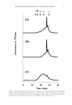
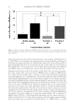
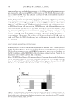
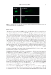
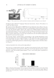
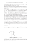
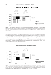
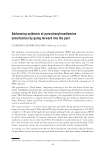
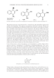
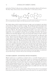
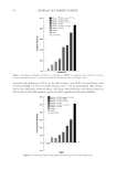
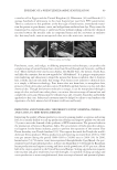
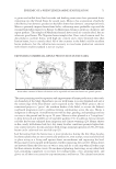
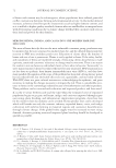
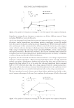
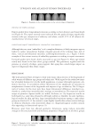
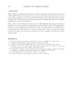
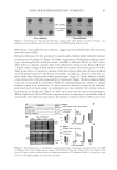
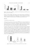
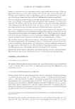
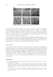
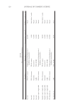
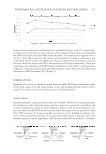
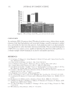
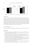
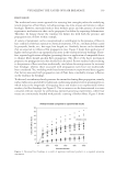
JOURNAL OF COSMETIC SCIENCE 88 the fi nal state of a dynamic system is the result of both the boundary conditions (includ- ing the initial conditions) and its dynamic characteristics, we could expect to have a more reproducible maturated tissue thanks to 3D bioprinting. SKIN EQUIVALENT STUDY DESIGN To reproduce complex, heterogeneous functional tissues and organs found in the human body such as skin, understanding of composition and organization of their components is an essential requirement. PREBIOPRINTING This is the fi rst step to generate a 3D tissue fi le containing the 3D structure and compo- sition of the tissue to be bioprinted. This is the product of an ideation phase based on the observation of native tissues or imaging data and literature analysis regarding dermal and epidermal histometry. ImageMatrix is used to generate complex visual designs for tissue. The goal is to determine and design specifi c virtual micropatterns to engineer at fi rst a dermis then an epidermis onto the dermis. Dermis design. As the native dermis in the skin is mainly composed of collagen I and III, we used both collagen type I and a mixture of collagen type I and III (95–5%) to associate with normal human adult fi broblasts in our preliminary testing. ▪ The 3D structure was created by alternating a layer of collagen with a layer of cells. ▪ The cell pattern was chosen to ensure a uniform distribution of fi broblasts. Figure 3. (A) Printed dermis after 5 days, (B) epidermized printed dermis (keratinocytes manually depos- ited) after 15 days, and (C) printed skin after 14 days. Figure 2. Laser-assisted bioprinter benefi ts compared to other bioprinters (6).
Purchased for the exclusive use of nofirst nolast (unknown)
From: SCC Media Library & Resource Center (library.scconline.org)