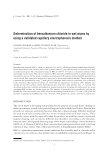
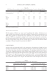
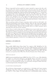
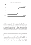
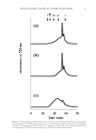
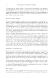
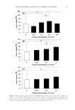
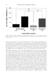
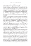
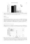
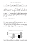
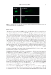
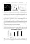
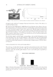
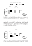
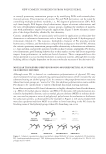
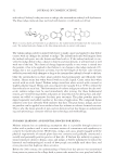
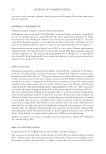
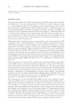
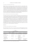
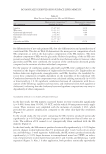
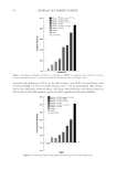
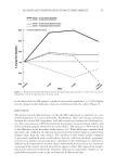
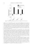
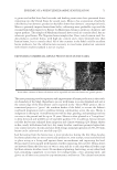
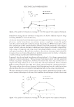
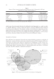
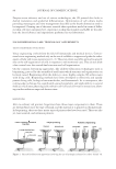
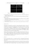
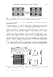
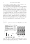
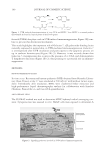
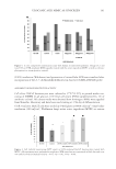
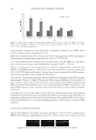
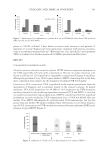
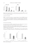
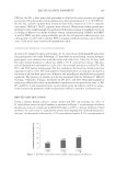
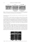
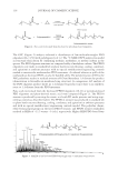
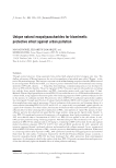
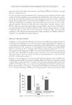
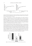
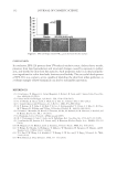
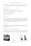
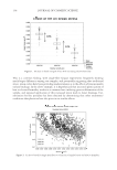
NEW BIOPRINTED SKIN, COSMETIC IN VITRO MODEL 89 ▪ In each plane, the cell pattern was a square network with a characteristic distance of 300 μm between spot of cells. ▪ Number of layers is designed to print a minimal initial dermis thickness of 200 μm. Epidermis design. ▪ The epidermis basal layer was designed with pattern was chosen to ensure a uniform distribution of normal human adult keratinocytes. ▪ In each plane, the cell pattern was a square network with a distance of 100–300 μm between spot of cells. ▪ Number of layers is designed to reach a minimal initial epidermal basal layer thickness. BIOFABRICATION Two different technologies were combined to 3D print the dermis equivalent, a microvalve technology was used to print the collagen layers, whereas LaBP was used for the cell layers. Culture media for fi broblasts is a DMEM/F12 base containing antibiotics and 20% SVF for seeding then 10% during growth period. Green medium (7) supplemented by 50 μg/ml ascorbic acid and antibiotics is used for keratinocyte seeding and DMEM/F12 supple- mented by (0.8% BSA, 0.12 UI/ml insulin, 0.4 μg/ml hydrocortisone, 50 μg/ml ascorbic acid and antibiotics) for keratinocytes differentiation. Kinetic of dermis maturation was performed to defi ne the best timing for dermis mat- uration before epidermal printing. One active ingredient (Origanum majorana leaf extract 0.04%—BASF) was added in the culture media to evaluate the benefi t on dermis matura- tion extracellular matrix synthesis, dermal–epidermal junction quality) and quality of epidermal anchorage and differentiation. ANALYSIS Dermis and epidermis quality was studied by histology and immunostaining. Results were compared to human skin biopsies and in house skin equivalent performed manually (Mimeskin®). Bioprinted skin models are suitable for dermocosmetic evaluations and allows to observe some changes induced by the treatment with an O. majorana extract used at 0.04% (in the dermis: LOXL1 elastin cross-linking enzyme—in the epidermis: thickness and involucrin). Figure 4. Immunostaining of untreated and treated models after 14 days.

JOURNAL OF COSMETIC SCIENCE 90 CONCLUSION We succeeded to develop a new evolution of 3D skin equivalent thanks to the optimization of 4D LaBP. This has now allowed us to produce a fully bioprinted skin that is close to current skin equivalents. It presents various advantages such as reproducibility and time for production. This model was used for the fi rst time to evaluate the effi cacy of a cosmetic ingredient (O. majorana leaf extract). Due to this recent advancement in the area of 3D bioprinted skin using laser-assisted bioprinter, we now anticipate the implementation of this model in the future with different cell types such as epidermal and/or AD stem cells in a specifi c pattern and defi ne micro- environment that will enable to be closer to human skin. Thus, we expect that these models will allow more predictive evaluation of active ingredient performance before clinical trials. REFERENCES (1) E. Bell, H. P. Ehrlich, D. J. Buttle, and T. Nakatsuji, Science, 211(4486), 1052–1054 (1981). (2) V. Yannas IV and J.F. Burke, J. Biomed. Mater. Res., 14(1), 65–81(1980). (3) S.T. Boyce, Christianson DJ, Hansbrough JF, J. Biomed. Mater. Res., 22(10), 939–957 (1988). (4) J. Malda, J. Visser, F. P. Melchels, T. Jüngst, W. E. Hennink, W, J. A. Dhert, J. Groll, and D. W., Hutmacher, Adv. Mater., 25(36), 5011–5028, (2013). (5) B. Guillotin, A. Souquet, S. Catros, M. Duocastella, B. Pippenger, S. Bellance, R. Bareille, M. Rémy, L. Bordenave, J. Amédée, and F. Guillemot, Biomaterials, 31(28), 7250–7256 (2010). (6) S. V. Murphy and A. Atala, Nat Biotechnol., 32(8), 773–785 (2014). (7) H. Green, O. Kehinde, and J. Thomas, Cell Biol., 11, 5665–5668 (1979).
Purchased for the exclusive use of nofirst nolast (unknown)
From: SCC Media Library & Resource Center (library.scconline.org)