232 JOURNAL OF COSMETIC SCIENCE
growing interest today in cosmetic products that come from naturally derived components.
Studies have screened abundant components from natural products, represented by arbutin,
tetrahydrocurcumin, and pterostilbene, all of which show favorable effects on pigmentation
reduction.5–7
Naturally whitening active ingredients can inhibit the formation and transfer of melanin
through multiple pathways.8 Many of these actives are reported to inhibit the transferring
of melanin from melanocytes to keratinocytes.9 For example, glabridin, a prenylated
isoflavonoid of G. glabra L. roots, has been associated with a wide range of biological
properties, including regulation of estrogenic, energy metabolism, neuroprotective,
and skin whitening in previous studies.10 The inhibition activity against tyrosinase and
melanin synthesis of glabridin may be the main whitening mechanisms.11–13 While the
present studies have mainly focused on the whitening effects of glabridin at the cell
level (melanocytes), few clinical reports on the effects of glabridin on melasma have been
published.
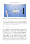
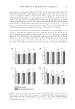
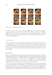
In this study, a skin 3D model was established and the effects of a whitening serum
containing glabridin on the apparent chromaticity, L* value, along with the content and
distribution of melanin were studied. Meanwhile, the whitening effect of the serum on the
melasma area and nonmelasma area was studied through clinical experiments.
MATERIALS AND METHODS
MATERIALS
Glabridin (98.5%) was bought from Push Bio-Technology (Push Bio-Technology, Ltd.,
Chengdu, China). Dimethyl sulfoxide and kojic acid were bought from Sigma-Aldrich
(Sigma, Massachusetts, USA). The 3D skin model provided was MelaKutis® (Guangdong
Boxi Biological Technology Co., Ltd., Guangdong, China). All other chemicals used were
of analytical grade.
PIGMENTATION IN 3D SKIN EQUIVALENT MODEL
The reconstructed human 3D epidermis (MelaKutis®) consisted of normal human-derived
epidermal keratinocytes and normal human epidermal melanocytes that had been cultured
to form a multilayered, highly differentiated model, similar to real skin structure. The blank
control (BC) group changed the culture medium every day without any other treatment
the negative control (NC) group, the positive control (PC) group with kojic acid (500 µg/
mL), and the whitening serum group were required to change the culture medium every
day and undergo ultraviolet-B (UVB) stimulation every day (50 mJ/cm2). The whitening
serum and PC groups used samples on the model surface on day 3 and day 5.The test
samples were applied topically in a volume of 10 µL. Controls were not treated. The change
in media and application of test material was repeated every 2 days for 7 days. The chemical
was added in the lower well of the reconstructed epidermis system then penetrated through
a membrane to reach the basal cells. The epidermis was then fixed with 4% formalin
in phosphate-buffered saline, followed by Fontana-Masson staining to visualize melanin
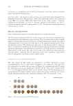
pigments. All tissues were photographed using a digital camera for a visual assessment of
depigmentation. It was then processed further for determining skin lightening as a measure
growing interest today in cosmetic products that come from naturally derived components.
Studies have screened abundant components from natural products, represented by arbutin,
tetrahydrocurcumin, and pterostilbene, all of which show favorable effects on pigmentation
reduction.5–7
Naturally whitening active ingredients can inhibit the formation and transfer of melanin
through multiple pathways.8 Many of these actives are reported to inhibit the transferring
of melanin from melanocytes to keratinocytes.9 For example, glabridin, a prenylated
isoflavonoid of G. glabra L. roots, has been associated with a wide range of biological
properties, including regulation of estrogenic, energy metabolism, neuroprotective,
and skin whitening in previous studies.10 The inhibition activity against tyrosinase and
melanin synthesis of glabridin may be the main whitening mechanisms.11–13 While the
present studies have mainly focused on the whitening effects of glabridin at the cell
level (melanocytes), few clinical reports on the effects of glabridin on melasma have been
published.
In this study, a skin 3D model was established and the effects of a whitening serum
containing glabridin on the apparent chromaticity, L* value, along with the content and
distribution of melanin were studied. Meanwhile, the whitening effect of the serum on the
melasma area and nonmelasma area was studied through clinical experiments.
MATERIALS AND METHODS
MATERIALS
Glabridin (98.5%) was bought from Push Bio-Technology (Push Bio-Technology, Ltd.,
Chengdu, China). Dimethyl sulfoxide and kojic acid were bought from Sigma-Aldrich
(Sigma, Massachusetts, USA). The 3D skin model provided was MelaKutis® (Guangdong
Boxi Biological Technology Co., Ltd., Guangdong, China). All other chemicals used were
of analytical grade.
PIGMENTATION IN 3D SKIN EQUIVALENT MODEL
The reconstructed human 3D epidermis (MelaKutis®) consisted of normal human-derived
epidermal keratinocytes and normal human epidermal melanocytes that had been cultured
to form a multilayered, highly differentiated model, similar to real skin structure. The blank
control (BC) group changed the culture medium every day without any other treatment
the negative control (NC) group, the positive control (PC) group with kojic acid (500 µg/
mL), and the whitening serum group were required to change the culture medium every
day and undergo ultraviolet-B (UVB) stimulation every day (50 mJ/cm2). The whitening
serum and PC groups used samples on the model surface on day 3 and day 5.The test
samples were applied topically in a volume of 10 µL. Controls were not treated. The change
in media and application of test material was repeated every 2 days for 7 days. The chemical
was added in the lower well of the reconstructed epidermis system then penetrated through
a membrane to reach the basal cells. The epidermis was then fixed with 4% formalin
in phosphate-buffered saline, followed by Fontana-Masson staining to visualize melanin
pigments. All tissues were photographed using a digital camera for a visual assessment of
depigmentation. It was then processed further for determining skin lightening as a measure