234 JOURNAL OF COSMETIC SCIENCE
15 minutes in a temperature of 20–24°C and humidity of 40–60% (relative humidity)
before instrumental measurements.
Statistical analysis. All statistical analyses of data were performed using OriginPro® 9.0
(OriginLab Corporation, Massachusetts, USA). The significance analysis was performed
using SPSS 13.0 software (IBM, New York, USA). In the analysis results, *indicates
comparison with baseline, p ≤ 0.05 **indicates comparison with baseline, p ≤ 0.01 #
indicates comparison with the control group, p ≤ 0.05 ##indicates comparison with the
control group, p ≤ 0.01.
RESULTS AND DISCUSSION
EFFECT OF WHITENING SERUM ON APPARENT CHROMATICITY OF MELANIN MODEL
The 3D reconstructed pigmented skin model composed of keratinocytes and melanocytes
was employed for skin whitening efficacy testing. The 3D melanin skin model is highly
similar to human skin structure and function. A multi-dimensional scientific detection of
whitening products was carried out with respects to the apparent chromaticity, L* value,
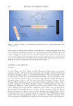
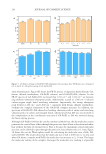
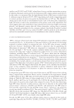
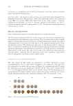
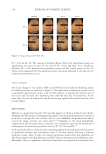
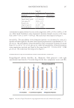
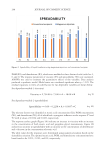
and melanin content.14 The chromaticity appearance of the melanin model is shown in
Figure 1. Compared to the BC group, the more darkened apparent chromaticity of the NC
group was significantly, while the apparent chromaticity of the PC group was significantly
white relative to NC group. The color of the model in the whitening serum group was
obviously lighter, indicating that the whitening serum had a significant improvement
effect on the apparent chromaticity of the melanin model.
CHANGES OF L* VALUE AND MELANIN CONTENT
The color change of skin model was measured by a L*a*b* chromaticity system
spectrophotometer. The L* value refers to brightness. The higher the value, the more
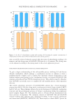
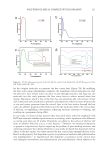
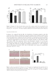
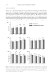
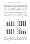
the color tends to be white, and vice versa. In Figure 2A, compared with the NC group,
the whitening serum group significantly improved the L* value of the melanin model
(P 0.01). Changes in melanin content are also shown in Figure 2B. The whitening serum
group displayed the lowest melanin content among the groups, indicating that treated
groups containing glabridin had a significant inhibitory effect on melanin (P 0.01).
Figure 1. Chromaticity appearance of the melanin model.
15 minutes in a temperature of 20–24°C and humidity of 40–60% (relative humidity)
before instrumental measurements.
Statistical analysis. All statistical analyses of data were performed using OriginPro® 9.0
(OriginLab Corporation, Massachusetts, USA). The significance analysis was performed
using SPSS 13.0 software (IBM, New York, USA). In the analysis results, *indicates
comparison with baseline, p ≤ 0.05 **indicates comparison with baseline, p ≤ 0.01 #
indicates comparison with the control group, p ≤ 0.05 ##indicates comparison with the
control group, p ≤ 0.01.
RESULTS AND DISCUSSION
EFFECT OF WHITENING SERUM ON APPARENT CHROMATICITY OF MELANIN MODEL
The 3D reconstructed pigmented skin model composed of keratinocytes and melanocytes
was employed for skin whitening efficacy testing. The 3D melanin skin model is highly
similar to human skin structure and function. A multi-dimensional scientific detection of
whitening products was carried out with respects to the apparent chromaticity, L* value,
and melanin content.14 The chromaticity appearance of the melanin model is shown in
Figure 1. Compared to the BC group, the more darkened apparent chromaticity of the NC
group was significantly, while the apparent chromaticity of the PC group was significantly
white relative to NC group. The color of the model in the whitening serum group was
obviously lighter, indicating that the whitening serum had a significant improvement
effect on the apparent chromaticity of the melanin model.
CHANGES OF L* VALUE AND MELANIN CONTENT
The color change of skin model was measured by a L*a*b* chromaticity system
spectrophotometer. The L* value refers to brightness. The higher the value, the more
the color tends to be white, and vice versa. In Figure 2A, compared with the NC group,
the whitening serum group significantly improved the L* value of the melanin model
(P 0.01). Changes in melanin content are also shown in Figure 2B. The whitening serum
group displayed the lowest melanin content among the groups, indicating that treated
groups containing glabridin had a significant inhibitory effect on melanin (P 0.01).
Figure 1. Chromaticity appearance of the melanin model.