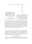
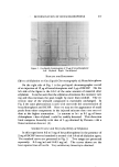
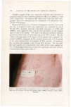
696 JOURNAL OF THE SOCIETY OF COSMETIC CHEMISTS Despite a substantial literature, certain unclear aspects of the nature of perfume-induced dermatitis prompted this work. As bergamot ap- peared to be the principal offender, it was decided to start with an in- vestigation of oil of bergamot and to identify those chemical components responsible for phototoxicity. This is a non-immunologic, light-induced skin response to a photoactive chemical likened to an exaggerated sun- burn. The fact that complainants sometimes used a perfume for a con- siderable time before suffering adverse skin effects suggested the possi- bility that allergenic or photoallergenic effects might also be involved (1, 2). This aspect, however, was not investigated here. Bergamot was first suspected of being capable of producing photo- toxicity by Freund, who reported in 1916 that four of his patients de- veloped erythema and pigmentation from eau de cologne containing bergamot (3). The term berlock dermatitis, spelled berloque-dermatitis in France and Berlockdermatitis in Germany (French breloque, German Betlocke ineaning pendant), has been given to the skin syndrome produced when bergamot oil, which is derived from the rind of Ciirus bergamia, an orange-like Mediterranean fruit, is applied to the skin and followed by exposure to sunlight. The name Berlockdermatitis or dermatitis in Betlock-Form (4) was coined by Rosenthal in 1924 (published 1925). Kuske (5) in 1938 appears to be the first to report that the furocoumarin components of plants are capable of producing photodermatitis. He isolated the furocoumarin bergapten from oil of bergamot and found it photoactive. In Germany, bergapten or 5-methoxypsoralen was known to be a component of oil of bergamot at least since 1839, when it was extracted from oil of bergamot by Ohme (6). In this work, often over- looked by investigators today, Ohme described the solubility character- istics and molecular weight of bergapten and correctly estimated the bergapten content of crude bergamot (0.37%). We now know that many plants contain photosensitizing furocoumarins (7). METHODS A llanovia "Inspectolite "* provided the radiation source in both buman and animal studies. The emission spectrum of one of these -- * H•novia lnspectolite with No. 16125, Type EH-4 bulb and red purple filter [Corning 7-39 (5874)]. Tfiis filter transmits 0% at 300 rim, 55% at 360 nm, and 0% at 410 nm as a more or less bell-shaped curve. It also has a frosted glass cut-off at 290 nm. Hanovia Izm H) Co., 100 Chestnut Street, Newark, N.J.

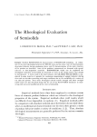
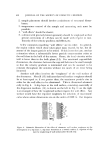
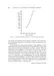
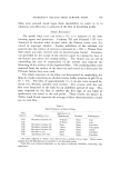
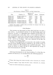
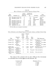
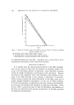
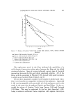
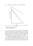
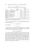
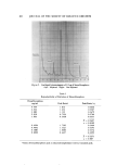
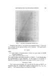
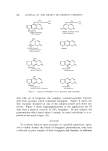
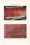
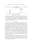
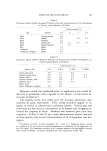
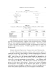
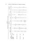
PERFUME PHOTOTOXICITY 697 radiation sources was •neasured* using a Perkin-Elmer monochromator with strip chart and tape recorders (8). Over 90t•o of the ultraviolet (UV) radiation output (between 300 and 400 nm) of the Inspectolite was found to be at 365 ----q- 5 nm. The total UV output was about 3000 /•watts/cm 2 at a distance of 10 cm from the source and 1900/•watts/cm 2 at 15 cm.* The first step was to isolate coumarins and [urocoumarins from bcrgamot and test them for phototoxicity on human subjects (Appendix I). As these first preparations were not entirely pure, this was con- sidered a screening step. Phototoxicity tests were then performed on nine subjects using oil of bergamot, the coumarin-psoralcn nonvolatile fraction, coumarin-psoralen components separated by thin-layer chro- matography (TLC), and an ethyl ether-precipitated portion of the cou- marin-psoralcns. Thus, in addition to oil of bergamot, which contains about 2% coumarin-psoralens, tests were performed on TLC band 1 con- sisting mainly of bcrgamottin, band 2 which was mainly 7-mcthoxy-5- geranoxycoumarin, band 3 which was about 96:4 limettin-bergapten, band 4 which contained elements of band 3 plus bergaptol, and an ethyl ether-precipitated portion which was recrystallized three times with methanol and was mainly bergapten. The chemical formulas are shown in Fig. 1. The human studies were performed according to the method of Burdick (10). The skin of the forearm was tape-stripped to glistening. The agent (0.05 ml) was applied to the stripped area (5 X 12 cm), allowed to remain undisturbed for 5 minutes, and then irradiated with the Inspectolite for 40 minutes at a distance of 8-10 cm. The arm was examined at 24 and 48 hours. RESULTS Results (Table I) show that phototoxicity, consisting of erythema, edema, and sometimes vesiculation, was easily obtained on stripped • Measuremcnts by Dr. M.P. Thekaehara, Goddard Space Flight Center, Greenbelt, Md. t Inverse square law is applicable if the source is assumed to be 8.2 cm behind the front of filter. The Inspectolite was calibrated with a G.E. Spectral Irradiance Standard lamp, a 1000-watt quartz-iodine lamp standardized by Eppley Laboratories, Newport, R.I. After the Inspectolite has been calibrated, it can be checked from time to time with a Jagger UV dose-rate meter fitted with a diaphragm over the photocell to reduce the UV output intensity (9). An important factor to note, especially in biologic experiments involving different exposure times, is that the output increases as the instrument warms up.
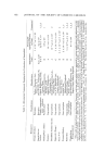
Purchased for the exclusive use of nofirst nolast (unknown)
From: SCC Media Library & Resource Center (library.scconline.org)