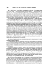
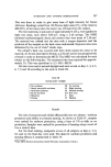
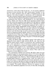
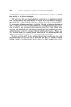
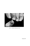
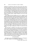
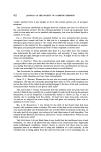
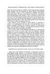
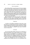
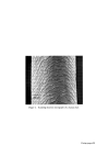
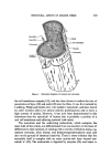
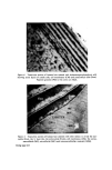
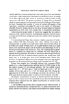
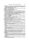
Figure 2. Scanning electron micrograph of a human hair. Facing page 430

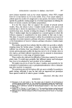
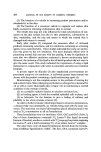
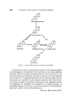
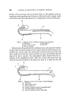
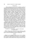
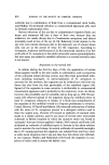
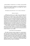
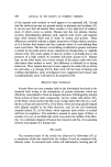
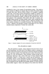
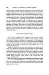
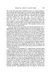
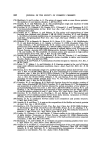
STRUCTURAL ASPECTS OF KERATIN FIBRES 431 Microfil Cuticle -- Cell membranes Nuclear remnants Figure 1. Schematic diagram of human hair structure. the cell membrane complex (18), and has been shown to reduce the rate of penetration of dyes (19) and acids (20) into the fibre. It was first isolated by Lindberg, Philip and Gral•n (21), who called it 'epicuticle', and later shown by other workers (22) to be almost entirely proteinaceous and to have a high content of cystine. However, it has now been demonstrated in this laboratory that the 'epicuticle' of human hair is probably a portion of a unit cell membrane and adhering material (vide infra). The exocuticle and the underlying endocuticle, which comprise the main bulk of the cuticle, are differentiated from one another on the basis of differences in their intensity of staining with a variety of electron stains, e.g. osmium tetroxide, silver nitrate, and dodecatungstophosphoric acid, and also on the ground of chemical reactivity. There is some evidence that the exocuticle itself is complex with an outer cystine-rich layer termed exo- cuticle 'a' (23). The endocuticle is digested by enzymes (24) and hence is
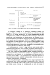
Purchased for the exclusive use of nofirst nolast (unknown)
From: SCC Media Library & Resource Center (library.scconline.org)