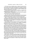
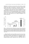
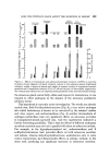
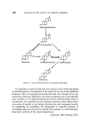
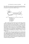
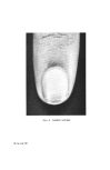
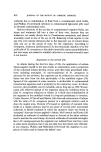
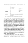
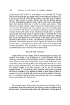
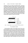
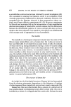
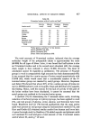
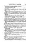
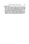
432 JOURNAL OF TIlE SOCIETY OF COSMETIC CHEMISTS considered to have a low content of cross-linking cystine. The cuticle is more resistant to diffusion of reagents than the cortex (25) and the shape of cuticle cells varies with the source of fibres. It is possible to separate cuticle cells, cortical cells, and the cell membrane complex by shaking wool fibres in formic acid (26). The acid rapidly disrupts and dissolves at least part of the cell membrane complex, setting free the individual cells. Amino acid analyses of the separated, whole cuticle show that it contains con- siderably larger amounts of cystine, proline, serine, valine, and glycine than the fibre as a whole (27-29). Cuticular protein is relatively amorphous and shows neither orientation nor crystalline formation. The schematic diagram of Fig. 3 indicates the generally accepted nomenclature for the various submicroscopic components of human hair cuticle cells. Hair surface 'a'LAYER -- Exocuticle Endocuticle Membrane (cell I) Cement or 8-band Membrane (cell 2) Portion of adjacenf cuticle cell Figure 3. Schematic diagram of the various components of human hair cuticle cells. The cell membrane complex The cell membrane complex, which originates from the fusion of two unit cell membranes, one from each of the adjacent cuticle or cortical cells, has been observed by electron microscopy of transverse sections by Rogers (30, 31) and other workers. The whole structure is about 30 nm thick and consists of a unit cell membrane, which probably contains protein and a bimolecular lipid layer, then a fairly thick layer of dense material called 'intercellular cement' or fi-band and a second unit cell membrane. The intercellular cement is easily digestible by trypsin (32) and its composition has been the subject of much speculation (33). The cell membrane complex may be extracted at least partially and possibly entirely by formic acid and certain milder reagents. Amino acid analyses of the extract show that this

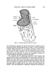
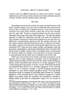
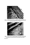
STRUCTURAL ASPECTS OF KERATIN FIBRES 433 protein is also very different from that of whole wool in that it contains smaller amounts of cystine, proline, threonine and serine and larger amounts of lysine, histidine and the aromatic amino acids (26). The cortex Proceeding inwards from the cuticle, the major structural feature of the fibre is reached, namely, the cortex. Horio and Kondo (34) noted that the cortex of fine wool fibres has a bilateral structure, in the sense that about one-half of the cortex always absorbs a given type of dye more intensely than the other half. Numerous reactions indicate that the more reactive side always lies on the convex side of the crimp wave. The side with greater reactivity is called the orthocortex and that with lesser the paracortex. This asymmetry between the orthocortex and paracortex has been shown in differences in morphological appearance (30, 35), staining behaviour (36) and certain chemical and physical properties (37). In bilateral wool fibres, the sulphur content of the paracortex is appreciably higher than that of the orthocortex (38). There are more cystine, proline, and glutamic acids but less glycine, phenylalanine, and tyro sine in the former than in the latter (28). The presence of a higher content of the cross-linking cystine would account for the lower degree of swelling of the paracortex, its greater resistance to acid hydrolysis and the slower rate of reduction of its cystine (39). Within each cortical cell of wool, which is about 80 •mlong and 5 •m in diameter, there is great complexity of structure. Electron micrographs of stained transverse sections of wool show that the cortex consists of approxi- mately circular macro fibrils (also referred to as 'tertiary aggregates' of the a-helices) having the appearance of whorls or spirals. Within the macrofibril are microfibrils (also referred to as 'secondary aggregates' of the a-helices) about 7.5 nm in diameter arranged in pseudo-hexagonal packing and embedded in a more heavily stained amorphous matrix.* The microfibrils themselves consist of a number of protofibrils (also referred to as 'primary aggregates' of the a-helices) about 2 nm in diameter arranged in a regular manner and packed within the micro fibril and embedded in intramicro- fibrillar matrix protein (40, 41). Independent support for the concept of *The electron-opaque appearance of structural units after fixation with metal compounds has been interpreted as an intense reaction of--SH or --S--S-- bonds with the metal, whereas an electron-translucent appearance is considered to be a weak reaction. Accordingly, the dense region seen in the cortex is thought to indicate the presence of amorphous protein(s) stabilized by numerous --S--S-- bonds, while the less dense region reveals the presence of a fibrous protein stabilized by less numerous --S--S-- bonds.
Purchased for the exclusive use of nofirst nolast (unknown)
From: SCC Media Library & Resource Center (library.scconline.org)