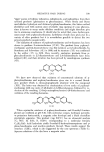
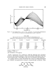
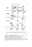
J. Soc. Cosmet. Chem., 24, 81-85 (February 2, 1973) The Histopathology of Wrinkles EDWIN T. WRIGHT, M.D., and WILLIAM V. R. SHELLOW, M.D.* Presented May 26, 1972, Seminar, Los Angeles, Calil •. SYNOPSIS-The HISTOPATHOLOGY of "WRINKLE SKIN" was compared to "non- wrinkle skin" with the patient acting as his own control. BIOPSY specimens were taken primarily from the FACE and NECK. Sections were fixed in formalin and stains were done for collagen, elastin, reticuhn, neutral and acid mucopolysaccharides, as well as the routine hematoxylin and eosin. It was not possible to differentiate wrinkle from nonwrinkle skin since the findings were similar in both areas. Wrinkles develop from body folds and from underlying muscle contraction. Older or aged skin and skin wltich has been damaged from sunlight is more subject to wrinkling. INTRODUCTION Wrinkling of the skin has been a source of concern among both sexes for centuries, and from the cosmetic standpoint vrinkles have been increasingly more important witness the phenomenal sales of hormonal face creams and the upsurge in the number of rhytidectomies and blepharoplasties being per- formed on both men and women in recent years. Although some knowledge is available concerning the .anatomical and structural changes involved in the formation of wrinkles, we have been unable to locate any previous studies dealing specifically with the histochemical alterations of wrinkles. The Formation o[ Wrinkles The superficial muscles of the face are those of expression, and they are inserted directly into the dermis. The muscle fibers diminish in size upon reaching the deep layer of the dermis, and they subsequently penetrate up- ward as fine fibers of the dermal papillae at the basal layer of the epidermis. The muscle fibers of these muscles of expression may contract independently, and thereby are capable of producing fine variations of expression. In child- hood, facial expression is less apparent, since the muscles are less well de- veloped and the subcutaneous layer of fat is thicker (1). * Veterans Administration, Wadsworth Hospital Center, Wilshire and Sawtelle Blvds., Los Angeles, Calif. 90073 81

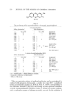
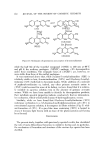
89. JOURNAL OF THE SOCIETY OF COSMETIC CHEMISTS Facial wrinkles are produced by repeated and habitual contraction of the underlying muscles of facial expression. When the facial muscles contract, the muscle shortens without a corresponding shortening of the overlying skin, thereby producing a wrinkle. The skin adapts itself by forming folds at right angles to the line of contraction of the underlying muscle (2). The supraorbi- tal wrinkle lines are caused by the contraction of the frontalis muscle which is inserted into the skin of the lower forehead. The transverse lines of the fore- head are due to the contraction of the frontalis muscle, and as the skin adapts itself to contraction of the muscle fibers, folds of excess skin are formed. In the upper eyelid, fine perpendicular strands of connective tissue terminate in the dermis to form the tarsal fold at the insertion of the levator palpebrae superioris. Similar tendinous insertions in the lower lid create the fine hori- zontal wrinkle lines which are accentuated by the contraction of the orbicu- laris oculi muscle (3). Other superficial facial muscles and their insertions are responsible for the other characteristic grooves and creases which are ap- parent in the face when it is not at rest. In later years, even when at rest, the face shows these lines and furrows quite well. Other factors, in addition to the pull of muscles, that result in the forma- tion of grooves and furrows which .are called wrinkles include the distribution and ratio of collagen and elastic fibers, the thickness of the skin and the amount of underlying fat, the water content of the skin, the activity of the appendegeal glands, and the biochemical changes of the connective tissue ground substance which occur with aging (4). Some histological studies of skin have shown that the process of aging is accompanied by a thinning of the epidermis with reduction in the size of the rete ridges. Volarelli (5) found that at the deepest portion of a wrinkle there was extreme thinning of the epidermis with only two or three layers of malpighian cells. The dermal papillae were completely absent, and the epi- dermal-dermal junction had become a straight line. On the other hand, Free- man et al. (6) did not find a statistically significant change with age in thickness of the epidermis of unexposed skin of the buttocks in individuals ranging in age from 25 to 76 years. With age, both collagen and elastic fibers undergo a change, most marked on exposed portions of the body. In exposed areas of the skin, .the fibers in the subpapillary layer show an increase in basophillic staining when hema- toxylin and eosin are used (7). The collagen fibers appear clumped and show basophilic degeneration. The elastic tissue also stains poorly, and has been described as being "fragmented." Examination of elastic ,tissue in thick sec- tions demonstrates that in areas of elastosis the fibers are increased in number and are tortuous, the latter accounting for the appearance of h'agmentation .as the microtome knife slices through the tissues (8). Unna, in 1896, described the alteration in connective tissue which is seen in aged (especially exposed) skin, and he felt that the sinfilarity of staining characteristics which devel- oped represented a fusion of collagen and elastin. Unna used the various
Purchased for the exclusive use of nofirst nolast (unknown)
From: SCC Media Library & Resource Center (library.scconline.org)