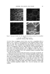
HISTOPATHOLOGY OF WRINKLES 83 descriptive terms, collacin, elacin, and collastin, •vhen discussing the altered connective tissue (9). Rather than entering into a detailed discussion of the complex and somewhat controversial changes which fake place in the corium as a function of aging, for our purpose the significant change is a loss of elasticity to the skin. The normal distensibility of the corium is due to the presence of the collagen fibers, although collagen itself is not elastic. The elastic fibers which stretch tend to prevent overextension of the collagen net- work (10). In aged skin, due to the alteration in both collagen and elastin, the normal elasticity of the skin is lost. Measurements of deformation of the skin in response to indentation sho•v that, while a decrease in normal elastic fibers correlates with the initial laxity of senile skin •vhen a deforming force is applied, as the force increases senile skin offers greater resistance than youthful skin (11). Increased intert•vining of collagen and decreased amounts of ground substance are felt to account for the reduced extensibility and in- creased tensile strength of elderly skin. Hydration is .a factor in •vrinkling, for the stratum corncure becomes de- hydrated .and brittle with aging, in large •neasure as a result of decreased wa•er-holding capacity (11). Some of the fine wrinkling seen in senile skin is attributable to this dryness of the horny layer, although the maior portion is due to the atrophy of the derreal papillae as discussed above. Also con- tributing to the decreased capacity of the skin to hold •vater is an involution of the sebaceous glands, and a reduction in the secretion of s•veat. Of the many and various changes which culminate in wrinkling, the altera- tions in ground substance appear to have especial importance to us. Such changes involve both the structural alterations of collagen and elastin and the permeability of the skin. Ma and Cowdry (12) noted a lack of clarity in the ground substance of senile skin •vhich was not apparent in the skin of younger individuals. Senile skin also shows an enhanced diffusibility of in- jected substances and a greater enhancement of spreading with the hyalu- ronidase (13). Smith et al. (14) noted a decrease in hexosamine, acid muco- polysaccharides, hyaluronic acid, and chondroitin sulfate with age. The changes in collagen which occur •vith age have been summarized by Gross (15). The fibril diameter increases with maturation but decreases slow- ly during senescense. There is an increase .of crystallinity. There is decreased swelling when collagen is subjected to dilute acids and there is a decrease in soluble collagen in acids. Resistance to collagenase is greater less base-bind- ing capacity, lower hexosamine-collagen ratio, higher hydroxproline content, and lower urinary hydroxyproline have been observed. MATERIALS AND METHODS Wrinkle specimens were obtained from 6 males and 6 females. The age range •vas 40 to 58 years. Wrinkle sites were primarily from the face, periorbi- tal region, forehead, neck, and, from one patient, the popliteal space. The

84 JOURNAL OF THE SOCIETY OF COSMETIC CHEMISTS wrinkles so obtained were fixed in formalin and were processed in the usual manner. Sections were cut at 8/• in a plane parallel to the long axis of the wrinkle, and also perpendicular to the long axis. Sections were stained with hematoxylineosin, Verhoeff's stain for elastic, periodic acid-Schiff reaction, alcian blue at pH 2.5, Mallory trichrome stain, and modified Wilders for reticulin, and were then examined under the light microscope. The surgical specimens contained adjacent normal skin making it possible to compare the vrinkle area with "nonwrinkled" skin. RESULTS The epidermis was slightly thinner at the base o[ the wrinkle than in the adjacent normal skin. This appeared to be due to compression of the epider- mal cells at the fold since the number of cell ]ayers was the same as in the adjacent normal skin. The size, shape, number, and staining characteristics of the elastic, reticulin, and collagen fibers were the same in the wrinkle and adjacent skin areas. An inconstant finding was slightly diminished neutral and acid mucopolysaccharides in the wrinkle area compared with the adjacent skin area. Skin specimens taken from light exposed areas revealed some basophilic degeneration of the collagen and an increase in elastic fibers consistent with sun damaged skin. DISCUSSION AND CONCLUSIONS It was not possible to differentiate "wrinkle skin" from "nonwrinkle skin" since the histopathology was similar in both areas. W'rink[es develop in body folds and from underlying muscle contraction. Skin previously damaged from sunlight, especially of the face and neck, and skin which is older (aged) is more subject to wrinkling. ACKNOWLEDGMENT The authors wish to acknowledge with thanks the kind cooperation Kurt Wagner, M.D., and Leonard Glass, M.D., in helping obtain wrinkle speci- mens for this study. (Received August 11, 1972) REFEltENCES (1) Kazanjian, V. H., and Converse, J. M., The Surgical Treatment of Facial Injuries, 2nd Ed., Williams and Wilkins Go., Baltimore, Md., 1959, p. 13. (2) Kraissl, C. J., and Conway, H., Excision of small tumors of the skin of the face with special reference to the wrinkle lines, Surgery, 9.5, 592-000 (1949). (3) Kazanjian, V. H., and Converse, J. M., op. cit., pp. 30-1. (4) Sobel, Harry, et al., The influence of age upon the hexosamine-collagen ratio of derreal biopsies from men, J. Gerontol., 13, 128-31 (1958). (5) Volarelli, F. F., Sulla Istologica e Sulla Patogenesi della Rughe, cited by Chieffl, M., Cowdry's Problems o[ Aging, 3rd ed., Williams and Wilkins Co., Baltimore, 1952, pp. 909-23.
Purchased for the exclusive use of nofirst nolast (unknown)
From: SCC Media Library & Resource Center (library.scconline.org)