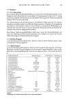
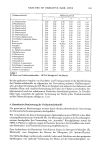
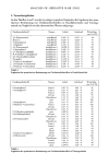
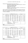
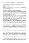
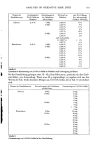
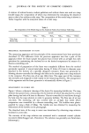
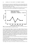
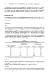
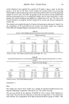
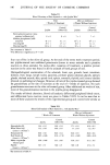
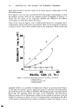
146 JOURNAL OF THE SOCIETY OF COSMETIC CHEMISTS Table VI Blood Chemistry of Rats Injected s.c. with Jojoba Wax a After 7 Weeks of Treatment After an Additional 6 Weeks Without Injections Control Control Jojoba (Olive Oil) Jojoba (Olive Oil) Serum glutamic-pyruvic trans- aminase (milliunits/ml) 8 _+ 0 8 _ 0 7 _+ 1 7 -- 1 Alkaline phosphatase (milli- units/ml) 45 + 6 41 + 3 56 b _+ 4 47 b -+ 3 Urea (mg/dl) 29 + 1 27 _+ 2 29 -+ 1 32 _+ 1 Glucose (mg/dl) 114 + 3 112 + 3 97 + 2 99 + 2 aSee comment in Table V •The difference is significant at P = 0.05 four out of five in the olive oil group. At the end of the seven week treatment period, ten jojoba-treated rats exhibited proteinuria (traces in seven animals and a positive reaction in three animals). Six weeks after cessation of treatment, a positive protein reaction in the urine was found in all the animals of both groups of rats. Histopathological examination of the adrenals, brain, eye, gonads, heart, intestines, kidneys, liver, lungs, lymph nodes, pancreas, prostate gland, pituitary glands, salivary glands, skeletal muscle, skin, spinal cord, spleen, stomach, thyroid, and urinary bladder showed no pathological changes. However, all rats of the jojoba-treated group showed fat granulomata at the site of injection at the end of 7 weeks of injection. No such granulomata were seen in the olive oil-treated group. After additional six weeks of rest, most of the granulomatous reaction in the jojoba group disappeared. The results of blood chemistry, blood cell analysis, differential peripheral blood count, and differential bone marrow count are given in Tables VI-IX. It can be seen that in most of these analyses the results of the experimental group and control were similar as Table VII Blood Cell Analysis of Rats Injected s.c. withJojoba Wax a After 7 Weeks of Treatment After an Additional 6 Weeks Without Injections Control Control Jojoba (Olive Oil) Jojoba (Olive Oil) Hemoglobin (%) 16.4 + 0.2 16.8 _+ 0.3 17.1 + 0.1 16.9 + 0.2 White blood cell 11.3 _+ 0.9 10.9 + 0.7 7.7 -+ 0.5 7.2 _+ 0.7 (count x 103/mm 3) Hematocrit (%) 49.0 + 0.6 49.0 + 0.4 50.0 _+ 0.5 49.0 + 0.6 Red blood cell 8.85 _+ 0.06 9.10 + 1.50 9.23 -+ 0.09 8.90 _+ 0.10 (count x 10•/mm 3) Mean corpuscular 56.0 _+ 0.06 55.5 + 0.8 55.0 + 0.5 55.2 _+ 0.4 volume (#m 3) Platelet 637 _+ 37 568 _+ 25 598 + 28 473 _+ 52 (count x 103/mm 3) aSee comment in Table V.

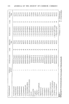
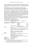
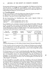
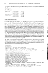
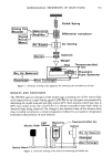
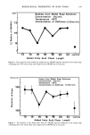
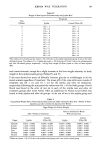
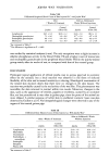
JOJOBA WAX TOLERATION 147 Table VIII Differential Peripheral Blood Count of Rats Injected S.C. withJojoba Wax a After 7 Weeks of Treatment After an Additional 6 Weeks Without Injections Control Control Jojoba (Olive Oil) Jojoba (Olive Oil) Lymphocytes 86.5 + 1.0 90.0 + 1.2 83.0 + 1.0 90.0 + 1.1 Neutrophilic granulocytes 7.2 + 0.6 5.0 + 0.8 9.7 b +_ 1.0 4.4 b + 0.9 Eosinophilic granulocytes 0.9 -+ 0.2 1.0 _+ 0.3 0.8 +_ 0.0 0.8 _+ 0.5 Monocytes 5.4 + 0.6 4.2 + 0.9 6.4 b + 0.6 3.2 • +_ 0.8 aSee comment in Table V. •The difference is significant at P = 0.005 was verified by statistical analyses (t-test). The only exceptions were a slight increase in alkaline phosphatase activity in the blood (Table VI) and a higher count of monocytes and neutrophilic granulocytes in the peripheral blood (Table VIII) in the jojoba-treated group mainly after six weeks of rest, as compared with that of the control group. DISCUSSION Prolonged topical application of refined jojoba wax to guinea pigs had no systemic effect on the animals, but a local reaction was observed in the form of reduced flexibility of the skin and increased sensitivity to shaving. Histological examination of the treated skin showed clearly that there were no changes in the skin tissues. The reaction was probably caused by the occlusivity of the wax film. This phenomenon was reversible the skin returned to normal within two weeks. Moreover, changes in the skin, such as the appearance of vesicles, papules or erythema, caused by an occlusive film, are less pronounced in man than in guinea pigs, since the pores of the animal are more dilated. A similar response of rabbit skin to undiluted cosmetic bases was also observed by Guillot et al. (7). No histopathological changes were observed in any of the organs of the treated guinea pigs. Table IX Differential Bone Marrow Count of Rats Injected s.c. WithJojoba Wax a After 7 Weeks of Treatment After an Additional 6 Weeks Without Injections Control Control Jojoba (Olive Oil) Jojoba (Olive Oil) Lymphocytes Myeloblasts Granulocytes Erythroblasts 4- normoblasts Eosinophils Basophils Plasma cells 24.8 + 1.8 21.7 + 2.2 23.4 + 1.6 26.8 +_ 2.6 22.5 + 0.7 25.4 + 1.8 20.5 + 0.7 20.0 + 0.6 22.6 + 1.1 22.3 + 0.4 23.9 + 1.4 23.1 + 1.4 26.5 + 1.8 27.1 + 3.1 28.4 + 1.6 26.8 + 3.0 1.7 + 0.1 2.4 + 0.5 2.3 + 0.3 2.1 + 0.5 1.4 + 0.2 1.3 + 0.3 0.6 + 0.2 1.2 + 0.4 0.3 + 0.1 0.2 + 0.1 0.5 +_ 0.1 0.5 +- 0.1 •See comment in Table V.
Purchased for the exclusive use of nofirst nolast (unknown)
From: SCC Media Library & Resource Center (library.scconline.org)