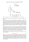
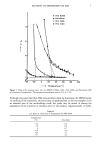
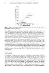
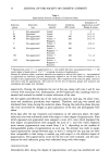
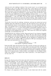
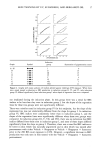
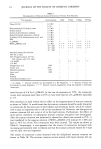
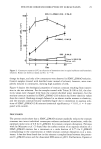
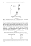
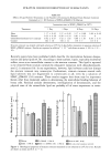
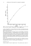
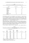
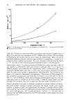
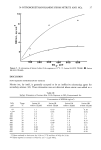
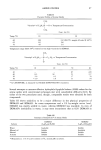
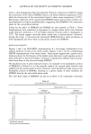
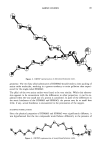
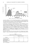
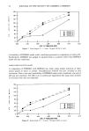
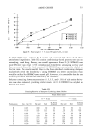
STRATUM CORNEUM DISRUPTION BY SURFACTANTS 23 denaturing agents (dimethyl sulfoxide and formic acid), the following keratolytic solutions were studied: saturated SA aqueous solution, 50% SA in ether (4), and a solution which contained 60% propylene glycol, 20% ethanol, 6% SA, and 14% distilled water (5). The effects of lipid depletion by ether, ethanol, and chloroform/methanol (2: 1) on the fragmentation of stratum corneum and on the counts of corneocytes released were exam- ined. Stratum corneum decomposition and corneocyte counts were assessed by the same methods described above. The rheological properties of the stratum corneum (20 x 5 mm) were measured using an extensometer (non-commercial instrument) at a constant extension rate (3.14 mm/min) while immersed in a test solution at 25øC for 2 hrs. Two mechanical parameters, viz. modulus of elasticity (tensional force/strain g/mm) and breaking strength (g), were obtained from the stress-strain curves. Each experiment was repeated four times. RESULTS The changes in stratum corneum sections immersed in test solutions (25øC) are pre- sented in Table I. In most of the surfactant solutions including Triton X-100 and SDS, the stratum corneum was not split into fragments, though rolling or curling occurred. Even in keratolytic and protein-denaturing agents, the stratum corneum was not re- duced to corneocytes. In sodium hydroxide solution the section of stratum corneum was split into many fragments. However, the fragments existed as large aggregates of corneocytes and could not be reduced further to individual intact corneocytes. On the other hand, the stratum corneum was gradually dispersed in C•2DMAO solution and finally disappeared into suspended individual corneocytes. It was comfirmed by micro- scopic observation that most of the stratum corneum cells in C•2DMAO solution re- tained their characteristic shape even after disruption to individual cells. The decompo- sition of stratum corneum was accelerated by addition of SDS to C•2DMAO solution. In a 0.1 M/1 solution of SDS/C•2DMAO (2:8) mixture, the stratum corneum was com- pletely reduced after 24 hrs immersion. However, the degree of stratum corneum de- composition was dependent on the concentration of the surfactant mixture, and it was not decomposed into fragments at less than the critical micelie concentration (ca. 5 x 10 - 4 M/l). Changes in the corneocyte counts with time of stratum corneum immersion in typical surfactant and keratolytic solutions are shown in Figure 2. Corneocytes were rarely observed in Triton X-100, SDS, and saturated aqueous SA solutions even after immer- sion for 30 days. In solutions of C•=DMAO, the number of corneocytes gradually in- creased with increasing immersion time. Corneocytes were counted in high numbers in the SDS/C•DMAO (2:8) mixture, and the count was almost constant with immersion time. These results corresponded to those from direct observation of the fragmentation of stratum corneum. However, in the case of sodium hydroxide solution there were almost no intact individual corneocytes in spite of the fragmentation of stratum cor- neum which occurred. Figure 3 shows the relationship between the corneocytes counted in aqueous solutions of SDS/C•DMAO mixtures (total concentration 0.1 M/l) and the mole fraction of C•DMAO. The corneocyte counts increased with increasing concentration of C•2 DMAO and then decreased after reaching a maximum. The maximum occurred at a

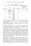
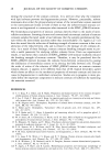
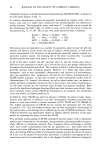
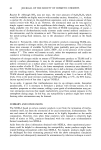
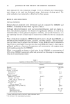
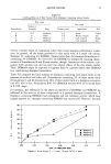
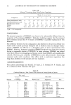
24 JOURNAL OF THE SOCIETY OF COSMETIC CHEMISTS Table I Decomposition of Stratum Corneum Sections in Various Test Solutions Test material Concentration i Day 3 Days 8 Days 30 Days Triton X-100 0.1 M/1 .... 0.1% .... Polyoxyethylene(15) dodecyl ether 0.1 M/1 .... Sodium laurate 0.1 M/1 .... Sodium dodecylbenzene sulfonate 0.1 M/1 .... Dodecyltrimethyl ammonium chloride 0.1 M/1 .... N-dodecyl N,N-dimethylamino acetic acid 0.1 M/1 .... SDS 0.1 M/1 .... C•2DMAO 0.1 M/1 4- 4- 4- 4- 4- 4- 4- 4- 4- SDS/C•2DMAO (2:8) 0.1 M/1 4- 4- 4- 4- 4- 4- 4- 4- 4- 4- 4- 4- 10 -2 M/1 4- + 4- + 4- 4- 4- 4- 4- 4- 4- 4- 10 -3 M/1 + + + + + + + + + + + + 10 -4 M/1 .... Saturated aqueous SA solution .... 50% SA in ether - - - 60% Propylene glycol, 6% SA, 20% ethanol, and 14% water .... 1 N NaOH + + + + + + + 1 H HC1 .... Thioglycollic acid 1 M/1 .... Formic acid 90% .... Dimethyl sulfoxide 1 M/1 .... Lactic acid 1 M/1 .... Ether .... Ethanol .... Chloroform/methanol (2:1) .... -: no change. +' Stratum corneum was decomposed to a few fragments. + +' Stratum corneum was decomposed to many fragments. 4- 4- 4-: Stratum corneum disappeared, being decomposed to individual corneocytes. mole fraction of 0.8 for C•2DMAO. In the case of immersion at 50øC, the corneocyte counts were increased more than at 25øC at every mole fraction of C•2DMAO, especially at 1.0. The immersion in lipid solvent had no effect on the fragmentation of stratum corneum as shown in Table I. It would seem that the stratum corneum should be easily disrupted to corneocytes by the extraction of intercellular and membrane lipids, which are consid- ered to play an important role in cell-to-cell attachment (6). However, the disruption of stratum corneum did not occur at all in the lipid solvents. Furthermore, the effects of lipid solvent treatment on subsequent stratum corneum disruption was investigated. After the stratum corneum was immersed in ethanol for a fixed time interval at 25øC, it was transferred to the aqueous solution of SDS/C•2DMAO (2:8) mixture (0.1 M/l) and kept at 50øC for 24 hrs. The corneocytes were then counted. The results are shown in Table II. Surprisingly, it was apparent that the counts of corneocytes decreased with increasing pretreatment time in ethanol and that the stratum corneum treated for 5 days was not split into any fragments. The results of corneocyte counts obtained with the delipidized stratum corneum are shown in Table III. The stratum corneum section treated with lipid solvents did not
Purchased for the exclusive use of nofirst nolast (unknown)
From: SCC Media Library & Resource Center (library.scconline.org)