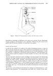
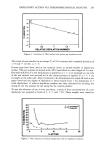
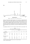
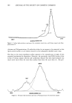
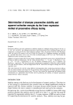
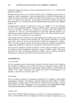
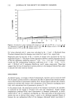
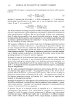
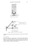
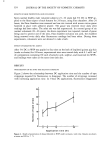
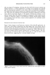
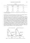
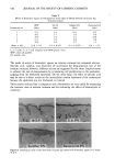
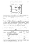
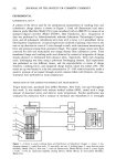
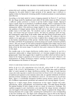
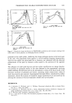
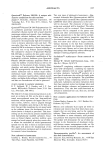
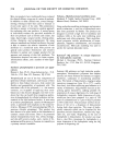
322 JOURNAL OF THE SOCIETY OF COSMETIC CHEMISTS This paper describes a more quantitative method to measure stratum corneum turnover and provides several examples to demonstrate its applicability. EXPERIMENTAL MATERIALS AND METHODS Dansyl chloride (DC) dispersed in white soft paraffin (WSP) was applied to the skin occlusively, using a Finn chamber (10-mm diameter). The depilation of guinea pig skin was accomplished using a commercial depilatory which contained wax and rosin. To confirm that the stratum corneum was completely stained by dansyl chloride, snip biopsies were obtained after staining. Frozen sections of skin tissue were prepared and examined under ultraviolet light using a Zeiss universal microscope. APPARATUS Fluorescence intensity was measured with a "split field" comparator (1) and a fluorom- eter (5). A comparator is described in a previous paper (1). It possesses a grey wedge that can be moved over half the field and a standard fluorescing strip. In use, the instrument is placed over the DC-treated test area of skin with the standard fluorescence adjacent to it, while both are illuminated by Wood's light. The grey wedge is adjusted to attenuate the brightness of the standard and to match it in intensity. The degree of fluorescence is recorded in arbitrary units on a scale attached to the grey wedge. Any results expressed by comparator units in this study were measured by this apparatus. Figure 1 shows a schematic drawing of a fluorometer, which contains a mercury lamp accompanied by a fluorescence body (Hamatsu Photonics Co., Ltd., L1549) at the center of a tube (3.5 x 17 cm). UV radiation with a peak at 338 nm is applied to the skin through an interference filter, F 1. The intensity of DC fluorescence is measured by a photomultiplier tube through an interference filter, F2. The fluorescence intensity depends on the nature of the incident UV radiation, but the UV-lamp specificity changes with time. Therefore, the strength of the DC fluorescence is expressed as a ratio of the incident ray strength. Output was calibrated with fluorescing paper before each experiment. PENETRATION OF DC INTO THE STRATUM CORNEUM Seven normal healthy subjects (5 male, 2 female), ages 24-56 years, were studied. Five per cent DC in WSP was applied under occlusion to the middle of the flexor aspect of the forearm for 3, 6, 12, and 24 hours, using a Finn chamber. The depth of DC penetration was examined by counting the number of adhesive tape strippings of stratum corneum required for the extinction of fluorescence. DC penetration was also examined by observing skin tissue taken by snip biopsy with a fluorescent microscope. MEASUREMENT OF TURNOVER TIME Fourteen normal healthy male volunteer subjects (21-46 years) had 5% DC in WSP applied to the flexor aspect of their forearm for 24 hours, using a Finn chamber. The fluorescence intensity at the application site and a nearby non-application site (control,

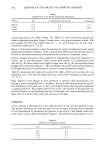
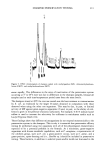
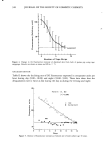
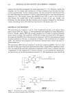
MEASURING TURNOVER TIME 323 --• F1 /Quartz glass Skin F2 I IS2 Fli12 Interference F,Iter Sl,S2 Photomultiplier Figure 1. Diagrams to show construction of the fiuorometer. I D•splay background) was measured daily. During this experiment, volunteers were not allowed to bathe. CIRCADIAN RHYTHM Seven normal healthy male volunteer subjects (21-36 years) were studied. After ap- plying 5% DC in WSP to the forearm (under occlusion) for 24 hours, the fluorescence intensity was measured twice daily (9:00- 10:00 in the morning and 5:00-6:00 in the evening) to examine the difference in desquamation rate during day and night. Volun- teers were not allowed to bathe during the experiment.
Purchased for the exclusive use of nofirst nolast (unknown)
From: SCC Media Library & Resource Center (library.scconline.org)