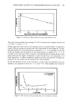
j. Soc. Cosmet. Chem., 38, 295-306 (September/October 1987) Adsorption of polymers and lipids on stratum corneum membranes as measured by ESCA E. D. GODDARD and W. C. HARRIS, Union Carbide Corporation, Specialty Chemicals Division, Tarrytown, NY (E.D.G.), and Bound Brook, NJ (W.C.H.). Received March i, 1987. Synopsis ESCA is shown to have the sensitivity to detect the presence of adsorbed conditioning polymers on isolated, treated stratum comeurn membranes and to rank them in terms of adsorbed amount. A cationic cellulosic polymer and chitosan show definite adsorption on the inner (dermis) side but less on the outer surface. This asymetric adsorption disappears upon pretreatment of the membranes with sodium dodecyl sulfate (SDS), thus implicating surface lipids in the observed deposition behavior. Conversely, a new, more hydrophobic cationic cellulosic polymer is found to adsorb symmetrically and to deposit to a greater extent than the other two polymers studied. This study also highlights the power of ESCA as a surface analytical tool. INTRODUCTION The barrier properties possessed by skin tissue depend to a large extent on the condition of the outermost skin surface, i.e., the stratum corneum. In addition, perceived ben- efits of the topical application of various conditioning agents can be related to the modification of the surface layer produced by these agents, and to their retention. Thus, it is desirable to have analytical techniques which are capable of directly examining the excised skin surface for the presence and concentration of such conditioners. Electron spectroscopy for chemical analysis (ESCA) is such a tool. The technique is inhe•rently surface-sensitive, providing chemical characterization of the outermost 25 to 50 A of the surface (1,2). Recently, ESCA has been applied to study the effects of various solvents and surfactants on lipid removal from stratum corneum membranes (3). The present study concerns the use of ESCA to examine the deposition of three syn- thetic polymers which are sometimes classified as conditioning agents on stratum cor- neum membranes. Two are cationic cellulosics and the third is a chitosan. Deposition on the outer and inner surfaces of the membranes is compared and the influence of prewashing the membranes with SDS on polymer deposition is reported. The ability of various emollients to provide protection from lipid removal by a surfac- rant wash is also examined using ESCA. 295

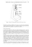
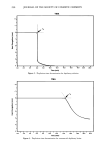
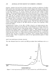
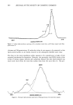
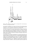
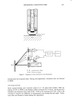
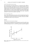
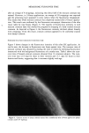
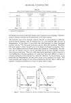
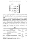
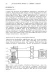
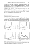
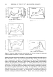
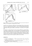
296 JOURNAL OF THE SOCIETY OF COSMETIC CHEMISTS EXPERIMENTAL MATERIALS AND SAMPLE PREPARATION The membranes were derived from neonatal rats obtained from Marland Farms using a procedure described previously (4). In brief, the excised skin samples were immersed in 60øC water for 30 seconds, and the stratum corneum membranes were carefully peeled away from the rest of the tissue. They were then air dried after careful flattening onto sheets of Teflon film. Throughout the procedure, care was taken to identify the outer surface and inner surface of the membranes. The two cationic cellulosic conditioning polymers used in this study, viz., UCARE Polymer JR 400 (CTFA designation Polyquaternium-10) and QUATRISOFT TM LM-200 Polymer (CTFA designation Polyquaternium-24), are products of Union Car- bide Corporation. The chitosan sample was of medium molecular weight, with a 1% aqueous solution viscosity of 2500 cps. Membranes were exposed to solutions of the polymers (0.1 weight % in distilled water) for 30 minutes with intermittent agitation, followed by three 30-second rinses in distilled water. The conditioned and rinsed mem- branes were air dried, as indicated above, on sheets of Teflon film. In cases where the membranes were pre-exposed to sodium dodecyl sulfate (SDS) solutions (1 weight % in distilled water), 30-minute exposures were used, again followed by three 30-second rinses. The emollients examined were castor oil, mineral oil (Arcoprime 350), oleic acid, gly- cerid acid (Agent G II, Westvaco), and lanolin. SDS treatment of membrane samples used in the emollient study was as noted above. Emollient treatments were performed at a level of 2.5 microliters/square centimeter applied to the outer skin surface, with the oils gently rubbed in for two minutes. The membranes were held in this condition for 30 minutes and then subjected to an SDS wash, rinse, and air drying, again under the conditions described above. ESCA ANALYSIS The membranes were analyzed as received using a Surface Science Laboratories Inc. SSX-100 ESCA spectrometer. The samples were mounted on a computer-controlled, rotating stage, using double-sided adhesive tape, such that no tape was present in the analysis area. The instrument is regularly calibrated to give the Au4f7/2 photoelectron peak at 83.93 eV and the Cu2p3/2 peak at 932.47 eV. An analysis area of approxi- mately 2 square millimeters was used throughout. All analyses were performed using an electron flood gun to provide charge compensation, and measured binding energies were charge-corrected to the principal C ls photoelectron line at 284.6 eV. Quantita- tion and peak fitting of high-resolution spectra were performed using software supplied by the manufacturer. RESULTS AND DISCUSSION CONTROL MEMBRANES AND THE EFFECT OF SDS WASHING Figure 1 is a representative survey spectrum of a sample of untreated control skin. Photoelectron peaks corresponding to carbon, oxygen, and nitrogen are readily ob-
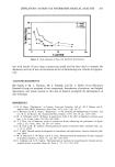
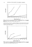
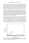
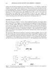
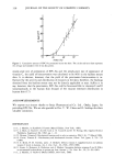
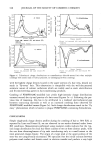
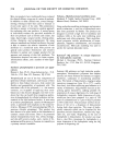
Purchased for the exclusive use of nofirst nolast (unknown)
From: SCC Media Library & Resource Center (library.scconline.org)