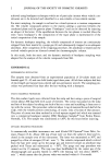
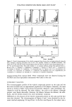
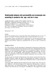
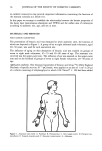
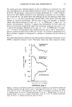
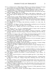
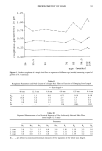
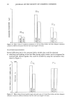
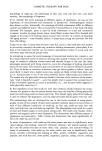
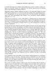
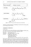
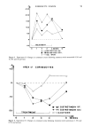
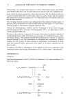
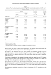
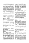
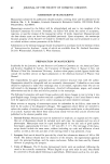
54 JOURNAL OF THE SOCIETY OF COSMETIC CHEMISTS Used Retinoids Chemical structure TRETINOIN I ._•C OOH ISOT RETINOIN OOH MOTRETINID ONHC2H 5 Figure 1. Chemical structure of the topical retinoids used in both experiments. surface area, corneocyte thickness, or morphology of corneocytes after removing them by the detergent scrub method (9). EXPERIMENTAL PROCEDURE MATERIALS USED The isotretinoin-acetone solution was kindly provided by Hoffmann-La Roche, Gren- zach, FRG, and motretinid by Wick Pharma, Grogg-Gerau, FRG. Motretinid = Monclerderma © gel 0.5% Isotretinoin = Roaccutan © capsules 2.5, 10, 20 mg Tretinoin = Eudyna © gel, cream 0.05% Airol © Cream, solution 0.05% Epi-Aberel © cream 0.5% VAS-Cordes © cream 0.05 % Retin-A © cream 0.1%, 0.05% gel 0.025%, 0.01% liquid 0.05% Two successive experiments were carried out. In the first study five healthy volunteers (four female, one male, ages 30-50 years) participated. The volar sides of forearms and upper lateral sides of arms were chosen as test sites. None of the subjects had any dermatological disorder at the time of investigation. In all experiments baseline deter- minations of corneocyte counts and areas were made. Each subject served at week zero as his own control (untreated skin). There was very little if any variance within the same

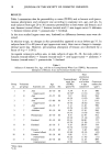
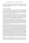
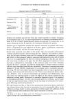
CYTOLOGY OF STRATUM CORNEUM 5 5 site when tested within a time period of six weeks (10). The following compounds, freshly made and used for not more than seven days, stored in a cool place and kept light protected, were applied: (1) 1.5% isotretinoin (dissolved in acetone) on the distal volar side of the right forearm (2) 0.05% tretinoin (dissolved in equal parts of ethanol and propyleneglycol) on the proximal volar side of the right forearm (3) ethanol and propyleneglycol on the distal upper part of the right arm (4) 0.5% motretinid on the distal volar side of the left forearm and (5) proximal to it, the gel base. Formulations were not randomized among application sites, as treatment response is negligible for small areas such as used in this study (unpublished data). The chemical structure of the retinoids are shown in Figure 1. Application was made daily for four weeks (five days per week). The compounds were applied to the skin with a cotton swab, so that a thin film covered the test area. Corneocytes were sampled once a week as described below. Two weeks after applications had been discontinued the last sample was obtained. In the second experiment another five volunteers (two female, three male, ages 26-32 years) took part. In this study two different concentrations of isotretinoin (1.5% and 0.5 %), dissolved in acetone, were applied on the right forearm. The solvent acetone was applied on the right upper arm. Two different concentrations of motretinid (0.5 % and 0.25%) and the gel base were tested on the left arm. Corneocytes were collected using the detergent scrub technique (1,9, 11). In this procedure a glass cylinder with an area of 3.8 cm 2 is pressed firmly onto the skin and 0.5 ml of phosphate-buffered 0.05% Triton-X-100 solution is added. The skin within the glass cylinder is rubbed gently with constant pressure with the blunt end of a teflon rod for 30 seconds. The suspension of corneocytes is obtained with an Eppendorf pipette. The corneocyte count per cm 2 skin surface increases with time of rubbing and pressure. Therefore, for all samples 30 seconds were kept constant (1,2). After staining with methylene blue and rhodamine B (2,9), some drops of the suspension were applied on a microscopic glass slide and cov- ered with a cover glass. The samples were evaluated 24 to 48 hours after they had been dried at room temperature. To count corneocytes the stained cell suspension was filled into a hemocytometer (Fuchs-Rosenthal) (1,2,9). The surface area of the corneocytes was analyzed using a projection microscope with a projection mirror (Carl Zeiss), a ) 100 oil immersion lens, and a semiautomatic analyzer system (Videoplan © Kontron, FRG). Fifty cells from each test site, randomly chosen, were evaluated and the mean value and standard deviation determined. The surface area was expressed in square mi- crometers (•m2). The error of this technique is less than 3% (12). RESULTS EXPERIMENT ! Clinical efficts. The visible effects caused by the retinoids on the skin were quite dif- ferent. To illustrate this we chose a classification system: (I) No visible irritation (II) slight irritation, i.e., some scaling (III)weak irritation, i.e., some erythema, scaling (IV) moderate irritation, i.e., erythema, scaling, some exudation and (V) strong irrita- tion with erythema, scaling, exudation, crusts. In the first experiment 1.5 % isotretinoin led to dermatitis in each test person (Table I). The volunteers reacted in a similar way to tretinoin. The vehicle (ethanol and propyl- eneglycol at equal parts) also caused some reactions probably due to the relatively high
Purchased for the exclusive use of nofirst nolast (unknown)
From: SCC Media Library & Resource Center (library.scconline.org)