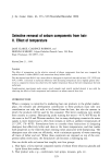
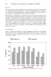
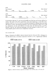
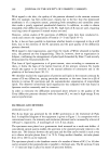
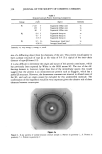
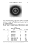
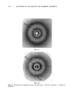
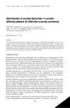
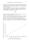
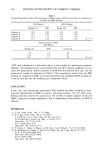
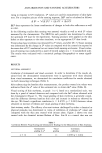
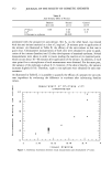
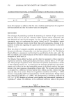
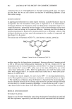
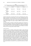
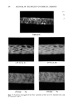
STRUCTURE OF STRATUM CORNEUM 353 Figure 3. Callus patterns in geometry//. By way of an example, distances of 62 and 43 fk were obtained with a specimen of breast skin. These distances are in good agreement with those observed by means of scanning electron microscopy in the human SC (8, 14). The bilayers would appear to be formed mainly of ceramides, as they only disappear following delipidation with a mix- ture of chloroform and methanol. The sharp equatorial reflections of group L2 and L3 would not originate from intercel- lular lipids, since they are derived from domains with a thickness of at least 0.3 Ixm. It Table II Stratum Corneum Lipid Components Group d (•) Aspect Intensity Solvent extraction L 1 62.7// Equatorial broad arc s Ch/met 45 // Equatorial broad arc s Ch/met 22.6// Equatorial broad arc s Ch/met 14 // Equatorial broad arc w Ch/met L2 42 // Equatorial sharp arc w Hexane 40 // Equatorial sharp arc s Hexane L3 33.5 l/ Equatorial sharp arc vs Ch/met 16.8// Equatorial sharp arc s Ch/met L4 6.2//ñ Meridianal sharp dotted ring vw Ch/met 5.8//ñ Meridianal sharp dotted ring s Ch/met 5.2//ñ Meridianal sharp dotted ring w Ch/met 4.6//ñ Meridianal reinforced ring m *Ch/met 4.1//ñ Meridianal reinforced ring vs *Ch/met 3.7//ñ Meridianal reinforced ring s *Ch/met Intensity: vs, very strong s, strong w, weak vw, very weak *not completely eliminated.

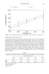
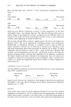
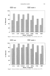
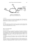
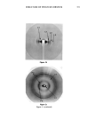
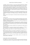
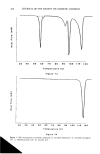
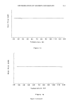
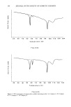
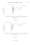
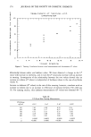
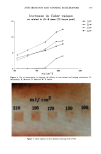
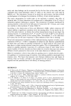
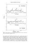
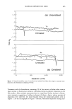
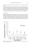
354 JOURNAL OF THE SOCIETY OF COSMETIC CHEMISTS P3 Figure 4a P3 P2 /./' Figure 4b Figure 4. X-ray patterns of delipidized stratum corneum sample: a. Pattern in geometry l b. Pattern in geometry//.
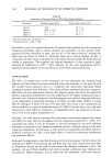
Purchased for the exclusive use of nofirst nolast (unknown)
From: SCC Media Library & Resource Center (library.scconline.org)