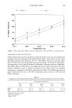
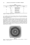
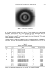
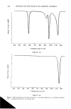
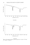
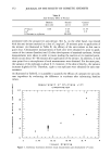
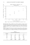
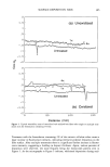
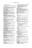
STRUCTURE OF STRATUM CORNEUM 355 is possible to infer the existence of a second class of lipids forming large crystallites located either within the corneocytes (corneocyte thickness range 0.3-0.7 •m, ref. 13) or inserted between the corneocytes but different from normal intercorneocyte lipids. Together with data published in the literature, our results (see below) show that choles- terol is responsible for the group L3 diffraction patterns (one harmonic of the arc at 33.5 •). Not all the cholesterol would thus be present in the intercorneocyte bilayers, but some would also segregate in crystals within the corneocytes or within certain dilated spaces between the cells. The group L2 patterns would be due to the crystallization of neutral lipids, since they disappear following hexane delipidation. Fatty acids or triglycerides, for example, may be involved, as they give rise to diffraction patterns in this domain of reticular dis- tances. Finally, group L4 would correspond to the distances between the hydrocarbon chains of the lipids. LIPOSOME STUDIES X-ray diffraction also allows the study of the molecular organization between lipids, surfactant molecules, and water (15). We therefore used this experimental set-up to characterize a suspension of liposomes formed from non-ionic lipids and cholesterol and stabilized by dicethyl phosphate (15). A 15 % solution was placed in very thin Lindeman glass tubes. A diagram characterized by a broad diffuse ring corresponding to a reticular distance around 53 ]t was obtained within a few minutes. The width of this ring is typical of thin vesicle walls (smaller than 5 bilayers). Transmission electron microscopy confirmed that the liposomes were indeed made of only a few bilayers. Following treatment of a specimen of human SC with this suspension, the diffraction pattern displayed two new features in the small angle part of the diagram: ß a group of sharp rings reinforced on the equator corresponding to a periodicity of 51 ß another family of sharp arcs on the equator corresponding to a spacing of 33.5 •. This latter phenomenon appears on the densitometric analysis of the patterns (Figure 5). It can be seen that the line at 33.5 • attributed to cholesterol is intensified and that the second harmonic that was absent in the control sample is clearly apparent in the treated sample. The problem of the actual location of these cristallites, inside or at the surface of the stratum corneum, remains open. CONCLUSIONS AND FUTURE PERSPECTIVES X-ray diffraction is a powerful means of obtaining a precise and quantitative description of supramolecular architecture in biological media. Applied to the study of the human

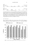
356 JOURNAL OF THE SOCIETY OF COSMETIC CHEMISTS Chol. (1) •' Control Tr Treated • Chol. (2) Unknown compone I I I I I I I 50 40 30 20 15 10 9 'r RETICULAR DISTANCE (.•.) Figure 5. Densitometric analysis of two patterns: liposome-treated stratum corneum and control. SC, this method had previously given only relatively poor quality images, as resolution was limited by the intensity of the X-ray beams on the one hand, and the low degree of crystallinity of the structure on the other. The use of the synchrotron overcomes these problems and the results presented here indicate the future importance of this technique for understanding not only the structure and function of the SC, but also the action of physicochemical systems used in modern day cosmetology. With regard to the first point, these results provide two new findings and confirm a previous report: The new findings concern our knowledge of the protein constituents of the SC. Contrary to what has been accepted for over 50 years, intercellular keratin in the normal SC would not appear to be in o• form. The main result leading to this interpretation is the total absence of any band at 5.1 • that corresponds to the pitch of the ot helix. Nonetheless, on the very dry SC of the callus of the hand, this band is clearly visible, suggesting that the keratinization process defines the supramolecular architecture of this protein this would have direct consequences on the binding of water molecules to the SC. Further studies of pathologic SC should provide new information in this field.
Purchased for the exclusive use of nofirst nolast (unknown)
From: SCC Media Library & Resource Center (library.scconline.org)