j. Soc. Cosmet. Chem., 43, 307-312 (November/December 1992) An in vitro method for screening sunscreen formulations for sun protection factor using a full-thickness skin model BARRY T. REECE, DAVID DEEDS, and MICHAEL ROZEN, Biophysical Skin Research Laboratory, Mary Kay Cosmetics, Inc., Dallas, TX 75247. Received April 14, 1992. Synopsis With the aid of a full-thickness skin model, minimum sun protection factors (SPF) of various sunscreens were determined. Sunscreen efficacy was determined by the release of interleukin IL- lot. The data show that this in vitro model is capable of determining minimum SPFs of a formula and may prove useful as a screen in the product development process. INTRODUCTION Sunscreens represent an important category of consumer products, permitting users to enjoy outdoor activities while reducing skin exposure to harmful ultraviolet (UV) light. In the United States, sunscreen products are regulated as over-the-counter drugs, and their safety and sun protection factor (SPF) claims must be established according to the Food and Drug Administration's regulations (1). These regulations require the SPF to be determined under specific conditions using human subjects. Historically, prior to final human SPF testing, in vivo SPF screening studies were performed using guinea pigs. With the suspension or banning of animal testing by many manufacturers of personal care products, a need now exists for reliable in vitro screening assays. Early attempts to define sunscreen protection by in vitro methods involved measuring the transmission of ultraviolet (UV) light through a dilute solution of sunscreen (2) or measuring the UV transmission through a film of sunscreen applied to a quartz glass (3). However, Groves et al. (4) were able to show that these methods cannot be used reliably due to their overestimation of the SPF value. Recently Diffey and Robson (5) described a rapid method for the determination of SPF by measuring the amount of ultraviolet light transmitted through Transpore TM tape with and without sunscreen applied close agreement between the in vitro and in vivo data was observed. Other investigators have measured the transmission of UV light through biological substrates (e.g., skin from hairless mice) as a means of determining SPF values (6,7). Keratinocytes are the principal cells of the skin that respond to UV irradiation, and their involvement in UV-induced inflammatory reactions has been established. Ansel et al. 307

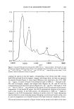
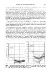
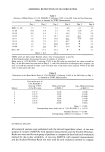
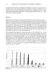
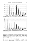
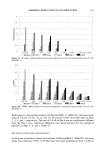
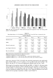
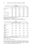
308 JOURNAL OF THE SOCIETY OF COSMETIC CHEMISTS (8) showed an increase in IL-lot production in keratinocytes exposed to UV irradiation, while Punnonen et al. (9) showed an induction of arachidonic acid after UVB irradia- tion. With recent application of cell and tissue culture assays as possible non-animal alternative safety and efficacy tests, we decided to determine if artificially grown human skin could provide a useful model system for screening SPF formulations. MATERIALS AND METHODS A three-dimensional artificial skin model, Skin 2 (Advanced Tissue Sciences, La Jolla, CA), was used to determine the SPF of various sunscreens. The artificial skin is com- posed of mitotically and metabolically active human dermal fibroblasts seeded onto a nylon mesh, while the multilayered epidermis is composed of a basal layer and several layers of differentiated and cornified keratinocytes. In order to assess the in vitro "sun protection factor", Skin 2 was placed into six-well tissue culture trays containing a Millicell TM insert (Millipore, Bedford, MA). The tray was covered by a 3.5" x 5" x ¾8" quartz glass plate to which was applied a 4-cm x 2.5-cm piece of Transpore TM tape (3M Company, St. Paul, MN). Sunscreens with SPF values ranging from 4 to 15 were applied to the tape at a concentration of 2 mg/cm 2. Additionally, sunscreen bases without sunscreen were tested. Skin was exposed to 42 mJ/cm 2 of UVA q- UVB radiation generated by a solar simulator (Solar Light Co., Philadelphia, PA). The port of the solar simulator was positioned 2 mm above the quartz glass to prevent removal of the product. The maximum time unprotected Skin 2 could be exposed to the UV radiation without releasing IL-lot was two minutes, which was defined as the in vitro minimal erythema dose (IVMED). Essentially, this is the in vitro equivalent of the human MED, which is based upon the time required for a fixed energy level of UV irradiation to elicit a visually discernible erythemic response in the skin of human subjects. Our background studies with the method (data not shown) has demonstrated that the Transpore Tape-Skin 2 system lacks the variability of human skin, and we are able to reproducibly observe two minutes as the IVMED. Accordingly, all sunscreen protection factors were multiplied by this factor to predict the exposure time. Once exposure was complete, the skin was incubated overnight at 37øC, 5% CO•. Following incubation, the media was assayed for the presence of IL-lot using an enzyme immunoassay kit (R & D Systems, Minneapolis, MN). Viability of the skin was deter- mined by the conversion of the dye 3[-4,5-dimethylthiazol-2-yl-]2,5,-diphenyltetra- zolium bromide (MTT) (Sigma, St. Louis, MO). RESULTS Figure 1 shows the effect of timed UV exposure vs skin responses (IL-lot release). No IL-lot was detected after two minutes of exposure however, by four minutes almost 8 pg/ml of IL-lot is present in the medium. Table I lists the results obtained from exposing skin to UV radiation with and without the application of sunscreen. In all cases where sunscreen was applied to Transpore tape affixed to the quartz glass, no IL-lot could be detected. In order to determine if IL-lot release was directly related to the
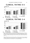
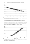
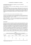
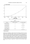
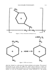
Purchased for the exclusive use of nofirst nolast (unknown)
From: SCC Media Library & Resource Center (library.scconline.org)