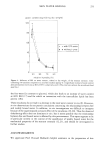
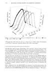
262 JOURNAL OF THE SOCIETY OF COSMETIC CHEMISTS rates across skin models, but receptor solutions with additives such as PEG-20 oleyl ether and cell culture medium improved their rate of permeation, although they did not alter skin concentrations (2). Recent investigations i, vitro on dermatomed human skin showed no evidence of increase of epidermal absorption or decrease of permeation from liposome-encapsulated trans- retinoic acid vs unencapsulated controls, but reported a decrease of permeation toward lower skin layers under extreme dose conditions (3). Previous work showed that nonionic surfactant vesicles had little or no direct effect on skin properties (4). Incorporation of salicylic acid into vesicles reduced its initial permeation into both hairless mouse skin and human skin i, vitro. After the preparation dried, there was a further decrease in permeation, suggesting a change of activity of the drug in the donor at the skin surface. The purpose of this study was to apply i, vitro permeation techniques to evaluation of skin uptake of RP from nonionic surfactant vesicles. One of the objectives was a comparison of permeation through excised hairless mouse skin as a substitute for human skin. Another goal was to assess if nonionic surfactant vesicles offer a controlled release system for topical delivery. MATERIALS AND METHODS CHEMICALS The receptor phases were surfactant solutions prepared with polyoxyethylene (20) iso- hexadecyl ether (Ariasolve TM 200, ICI Specialties, Wilmington, DE), polysorbate 80 (Tween © 80, ICI Specialties), and hydroxypropyl beta cyclodextrin (American Maize- Products Company, Hammond, IN). Retinyl palmitate, ascrobyl palmitate, and vita- min E (all from Hoffmann-La Roche Inc., Nutley, NJ) were used without any other purification. Light mineral oil was purchased from Fisher Scientific, Fair Lawn, NJ. All other chemicals were reagent grade. Retinyl-T2 5H palmirate, 0.1 mCi/g, was obtained from Hoffmann-La Roche. MANUFACTURE OF NONIONIC SURFACTANT VESICLES Neutral vesicles were prepared, according to a patented method (6), from polyoxyeth- ylene (2) cetyl ether, 72%, and cholesterol, 28%. Positively charged vesicles contained 6.2% dimethyl dialkyl (C2o-C22) ammonium chloride, while negatively charged vesi- cles contained 3.1% oleic acid. Ratios of the other ingredients were kept constant. The final pH of the preparation was measured at pH 5.5. The manufacture of nonionic surfactant vesicles (NSV) had to be modified to take into account the sensitivity of RP to oxygen, heat, and light. Manufacture was done in the nitrogen environment of a glove-box using tinted glass containers. The lipid phase mixture was heated above 60øC, introduced in syringe 1, and brought to 40øC. A measured quantity of RP was introduced in syringe 2. The two syringes were connected, and the preparation was transferred continuously from syringe 1 to syringe 2 to mix the two phases. When the mixture was contained in syringe 2, syringe 1 was disconnected and replaced by syringe 3 containing a measured quantity of deionized water at 40øC.

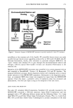
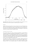
SKIN PERMEATION FROM VESICLES 263 The mixing operation was then repeated until a smooth preparation was achieved. The preparation was then introduced in a tinted glass container and used within 48 hours. STABILITY OF NSV Physical stability had to be achieved in order to obtain a reproducible and usable product. A 5-ml sample of the preparation was introduced in a tube and centrifuged at 6170 g for one hour. At the end of the centrifugation, the tube was inspected for phase separation and RP separation. Phase separation was identified by a separation of a clear water layer in the tube, and RP separation was identified by yellow droplets at the surface of the preparation. Chemical stability was optimized by using preservative system (methyl paraben 0.15 %, propyl paraben 0.15%) and antioxidant system (ascorbyl palmitate 0.1%, dl-alpha tocopherol 0.1%). The pH was 5.5 at the end of the manufacture. SKIN Hairless mice were purchased from Charles River (Wilmington, MA). Animals were killed and the skin removed. Dermatomed excised human skin (approximately 300-•m thickness) was purchased from the National Disease Research Institute (NDRI, Phila- delphia, PA). The sample was an abdominal skin sample (NDRI No. 25194) shipped frozen in dry ice. The skin was inspected visually and tested for damage using tritiated water. The flux obtained were compared to literature data (6). No damage could be detected visually and analytically. PERMEATION APPARATUS The apparatus used was a semi-automated system purchased from Crown Glass. The diffusion cells were made of Teflon, and the receptor side was of a flow-through design. Receptor fluid circulated through and was collected in vials. The cell was maintained at 32øC by a water-circulating system. The cell surface was 0.636 cm 2. The apparatus consisted of the cells, a fraction collector, a peristaltic pump, a cell heater, and a heating circulator. The receptor phase (saline solution containing 0. 125% chlorobutanol plus 3% surfactant) was pumped through the cell at a flow rate of 6 ml/hour, ensuring sink conditions during the duration of the experiment. This phase was collected at one-hour time intervals in scintillation vials. For a finite dose experiment, the donor compartment was open to the atmosphere and the experiment was run for 24 hours. All permeation experiments were run in triplicate (n = 3). SCINTILLATION COUNTING The liquid scintillation cocktail (Scintiverse TM BOA Scintanalyzed TM, Fisher Scientific, Fair Lawn, NJ) was added, and the radioactivity in the vials was counted for a ten- minute period to obtain a precise estimate of the radiolabeled 3H RP permeated (Beck- man LS 5000 TD, Beckman Instruments, Fullerton, CA). Previously a quench curve
Purchased for the exclusive use of nofirst nolast (unknown)
From: SCC Media Library & Resource Center (library.scconline.org)