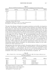
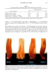
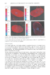
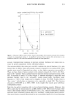
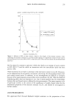
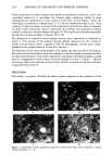
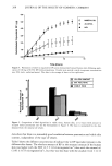
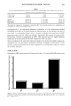
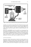
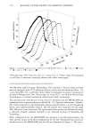
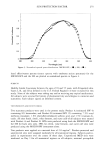
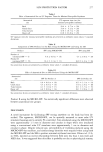
278 JOURNAL OF THE SOCIETY OF COSMETIC CHEMISTS instead, it may be explained by edge scattering or blurring afforded under the template configuration used to control site size in the investigation. This edge effect may have caused difficulty in subsequent visual perception of erythema. Optical fiber delivery of UV energy, as provided by the MICRO-SPF, more closely simulates the configuration of conventional UV delivery systems and thus circumvents the edge effect linked to Olson's work. Several advantages in experiment execution were provided by the MICRO-SPF com- pared to the SSS. Reduced site size lessened the area of insult for subjects, and simul- taneous irradiation of complete dose series reduced total experiment time. Additionally, randomization of microfibers at the common end of the 5-furcated optical fiber bundle facilitated uniform distribution of the UV energy among delivery fibers (Figure 2) and elimination of "hot spots." Conventional solar UV simulators commonly possess "hot spots" within the cross section of energy delivered to the skin these lead to non-uniform irradiation of subsites and subsequent difficulty in perception of erythema. Incident UV energy was collimated upon exit from delivery optical fibers of the MICRO-SPF thus, variability of energy intensity due to fiber distance from skin level was greatly reduced. Cripps (15) reported similar design modifications (collimated light) to better simulate sunlight. Conventional solar UV simulators deliver nearly collimated energy that focuses at a prescribed distance from the source housing this can lead to result variability since distance from source housing to skin level is rarely maintained constant over extended dose-delivery periods. Filter configurations for the MICRO-SPF and the SSS were designed to create close overlap of output spectra (Figure 3) and erythema effectiveness spectra (Figure 4). Since SPF efficiency is a function of the spectral transmission characteristics of the test sun- screen and the erythema effectiveness spectrum of the radiation source, comparability of SPF results produced using various radiation sources relies heavily on the relative same- ness of the spectra produced by the different sources. Sayre and Poh Agin (16) used hairless mouse epidermis in vitro to predict differences in SPF efficiencies based on UV radiation sources with differing spectral characteristics. LeVee eta/. (17), Noda eta/. (18), and Fukuda eta/. (19) reported higher SPF efficiencies using radiation sources rich in short-wave UVB and/or UVC compared to SPFs produced using radiation sources void of these wavelengths our own data (not published) con- firmed these results. In the present study, no statistical differences ((x = 0.05) were found between SPFs produced using the MICRO-SPF and SPFs produced using the SSS. These results suggest that differences between the radiation source output of the MICRO-SPF and the output of the SSS were small and, consequently, had no effect on product performance. Time for MPE for each anatomical site was statistically different (p 0.0001) from all other sites average times were 29.4 sec for back, 33.8 sec for face, 47.8 sec for volar forearm, and 61.8 sec for outer calf. The fact that MED is a function of anatomical position is well established. Olson et al. (8) reported MEDs differed by as much as fourfold, depending on body site. Although MEDs were assessed eight hours after irradiation, results of the study support our findings: trunk and head sites were found to be more sensitive than arm and leg sites arm sites were more sensitive than leg sites. Rhodes and Friedmann (10) found 24-hour MEDs to be significantly higher for buttock skin compared to back skin. Farr and Diffey (9) showed considerable MED variability

SUN PROTECTION FACTOR 279 over the back with lower-back sites higher for all subjects. The results of these studies support our findings that natural photoprotection differences exist as a function of anatomical site and that the back possesses the greatest UV sensitivity. No statistical differences (or = 0.05) were found between SPFs produced on various anatomical sites. Kawada et al. (20) reported that SPF efficiency decreased with increas- ing MED however, these data were produced on a single anatomical region. Fukuda et al. (19) demonstrated that SPF efficiency was a function of skin color and showed that lighter-skinned individuals produced higher SPFs. Although the results of numerous investigators (2-7) suggest that physiological differences in skin may affect sunscreen film formation, and thus sunscreen efficiency, the results of the present study suggest that SPF efficiency for products tested was not a function of anatomical site. Results of the present study demonstrate consistency between SPFs produced using the MICRO-SPF and SPFs produced using the current TFM method. More importantly, the results establish the effectiveness of the MICRO-SPF instrument for facilitating exam- ination of sunscreen efficiency on any anatomical region while using much lower total doses of UV energy compared to traditional systems. Currently FDA-proposed sunscreen testing guidelines mandate irradiation of test subsites no smaller than 1 square centi- meter (1), which would exclude use of the MICRO-SPF for final monograph testing. However, this limitation has been formally addressed via a proposal to the FDA for modification of the monograph for inclusion of devices such as the MICRO-SPF in the final monograph. Regardless, the MICRO-SPF instrument provides a means for efficient screening of photoprotection products. REFERENCES (1) (2) (3) (4) (5) (6) (7) (8) (9) (10) (11) (12) US Department of Health and Human Services, Sunscreen drug products for over-the-counter human use tentative final monograph, Federal Register, 58(90), 28194-28302 (1993). E. Tur, M. Tur, H. I. Maibach, and R. H. Guy, Basal perfusion of the cutaneous microcirculation: Measurements as a function of artatomic position. J. Invest. Dermatol., 81(5), 442•i46 (1983). International Commission on Radiological Protection, Report of the Task Group on ReJ•rence Man (Per- gamon Press, New York, 1974), pp. 46-50. A. B. Cua, K.-P. Wilhelm, and H. I. Maibach, Elastic properties of human skin: Relation to age, sex, and anatomical region, Arch. Dermatol. Res., 282, 283-288 (1990). S. Makki, J. C. Barbend, and P. Agache, A quantitative method for the assessment of the micro- topography of human skin, Acta Dermatovener (Stockholm), 59, 285-291 (1979). J. Hatzis, Skin surface profile technique and its applications, Int. J. Cosmet., 13, 281-291 (1991). L. Emtestam and S. Oilmar, Electrical impedance index in human skin: Measurements after occlusion, in 5 anatomical regions and in mild irritant contact dermatitis, Contact Dermatitis, 28, 104-108 (1993). R. L. Olson and R. M. Sayre, Effect of anatomic location and time on ultraviolet erythema, Arch. Dermatol., 93, 211-215 (1966). P.M. Farr and B. L. Diffey, Quantitative studies on cutaneous erythema induced by ultraviolet radiation, Br. J. Dermatol., 111, 673-682 (1984). L. E. Rhodes and P.S. Friedman, A comparison of the ultraviolet B-induced erythemal response of back and buttock skin, Photodermatol. Photoimmunol. Photoreed., 9(2), 48-51 (1992). R. L. Olson, J. Gaylor, and M. A. Everett, Skin color, melanin, and erythema, Arch. Dermatol. 108, 541-544 (1973). W. Westerhof, O. Estevez-Uscanga, J. Meens, A. Kammeyer, M. Durocq, and I. Cario, The relationship between constitutional skin color and photosensitivity estimated from UV-induced ery- thema and pigmentation dose-response curves, J. Invest. Dermatol., 94(6), 812-816 (1990).
Purchased for the exclusive use of nofirst nolast (unknown)
From: SCC Media Library & Resource Center (library.scconline.org)