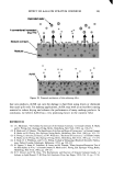
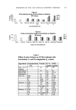
SKIN PENETRATION PROPERTIES 151 Adhesive tape stripping. Stripping (Tesa © 4204, BDF, Hamburg) was used to remove the outermost layers of the stratum corneum in sequence about ten layers of horny-layer cells (roughly 10 l•m) were removed. The mass of the removed horny-layer cells was constant for all layers, being approx. 1.10 mg per adhesive strip (1.9'10 cm), with a standard deviation of +0.36 mg (n = 20). The dermatome sections parallel to the skin surface were cut into 20-pm-thin sections and went down 200 pm through the epider- mis into the upper layer of the dermis. Detection of the vitamins. The detection of vitamin E, vitamin E acetate, and D-panthenol in the adhesive strips from the outermost skin layer and in the dermatome sections of the dermis and epidermis was based on extraction of the analytes and subsequent liquid chromatographic analysis of the obtained extracts. The selective determination of the vitamins in the complex matrix required substance-specific detection to be carried out after the chromatographic separation. The quantification was based on external standard calibrations and comparison of the treated and untreated skin. Detection of vitamin E and vitamin E acetate. Vitamin E/vitamin E acetate were extracted from the relevant skin sections with ethanol, and then the solvent was quantitatively removed and the residue was taken up in hexane. Extraction from adhesive strips was carried out directly with hexane. Vitamin E was separated from unwanted accompanying substances in a diol phase (Lichrospher 100 Diol) by means of liquid chromatography. The chromatography was carried out under isocratic conditions with n-hexane/te•-t. butyl methyl ether as eluent. Selective detection was performed with a fluorescence detector. The chromophoric system in the vitamin E molecule enabled the work to be carried out at an excitation wavelength of 295 nm. Detection ofpanthenol. In contrast to vitamin E and vitamin E acetate, panthenol has no molecular properties that would facilitate sensitive and selective detection in difficult matrices. After extraction from the skin or adhesive strips, therefore, the obtained extract was first hydrolyzed alcoholically and the panthenol quantitatively converted to ami- nopropanol. This hydrolysis product was separated from unwanted accompanying sub- stances in an ion-exchange column with diluted sodium hydroxide solution as eluent, and was converted to a strongly fiuorescing isoindole derivative in a postcolumn reaction with orthophthaldialdehyde. The fluorescence emission was measured with an HPLC fluorescence detector at a wavelength of 455 nm. RESULTS AND DISCUSSION PHASE BEHAVIOR OF THE COSMETIC FORMULATIONS For the leave-on products, consideration also has to be given to their behavior after open topical application. When they are applied, the emulsions are spread over the skin, forming a film (approx. 400 l•m). As the temperature of the emulsions increases to skin temperature, components with a high vapor pressure, in our case water, start to evaporate (3,4). Drying experiments on thin emulsion films show that•clepending on atmo- spheric humidity and the thickness of the layer--almost all the water escapes from the emulsions within five to ten minutes. During this period the viscosity and sometimes also the structure of the emulsion change (10).

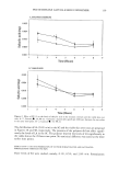
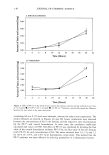
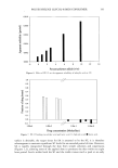
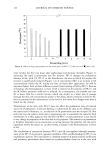
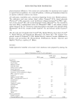
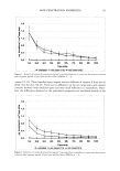
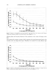
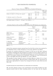
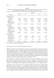
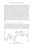
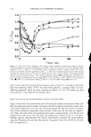
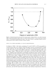
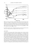
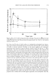
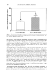
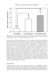
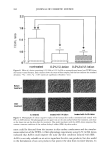
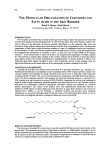
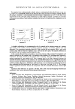
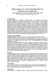
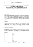
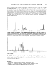
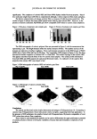
152 JOURNAL OF COSMETIC SCIENCE Directly after application, the lameliar cream contains 70% water and has a moderate viscosity of about 1000 mPas (Table I). When water evaporates, the lameliar layers are packed together more densely, leading to an appreciable viscosity increase (about 7,000 mPas). When the water content becomes less than about 15%, the amount of water is insufficient to allow o/w-emulsions to be formed any longer, and w/o-emulsions are formed (10). The situation is different with the w/o-cream. Here the oil is the outer phase and the viscosity rises exponentially by increasing the proportion of the inner water phase (10). During drying, therefore, the viscosity of the w/o-cream decreases below 10 mPas, in contrast to the o/w-emulsion. For the shower gel, the phase behavior will not change during the short application period. The surfactant product is washed away with a surplus of water after only two minutes. Therefore, phase changes caused by water loss will not take place under rinse-off conditions. KINETICS OF VITAMIN PENETRATION INTO THE SKIN UNDER LEAVE-ON CONDITIONS After application on the skin, an active ingredient begins to diffuse out of the galenic vehicle into the top layers of the skin. If, for purposes of simplification, it is assumed that the horny layer is a homogeneous diffusion barrier, then, according to Fick's law (Equa- tion 1), the penetration rate after a steady state has been reached depends only on the mobility of the active-ingredient molecule, given by the diffusion constant D, the coefficient of distribution K of the active ingredient between the vehicle and the stratum corneum layer, and the concentration c of the active ingredient (1,2). Penetration rate J = K/d ß D * c (Eq. 1) where d is the thickness of the horny layer, D is the diffusion constant, c is the concentration, and K is the distribution coefficient. Figures 1 to 4 show the penetration profiles of vitamin E and D-panthenol into the stratum corneum from w/o and o/w creams. The amount of naturally occurring vitamin E in the skin is several orders of magnitude smaller than the amount absorbed after topical application of the creams. For the oil-soluble vitamin E, a steep concentration gradient over the stratum corneum is observed (Figures 1 and 2), which indicates that the stratum corneum is the main penetration barrier. In contrast to the lameliar o/w emulsion, for the w/o emulsion there is no difference in penetration between 1 hour and 5 hours. This means that a steady state is already reached for the vitamin E penetration from the w/o emulsion after 1 hour. In general, the penetration is faster and much higher from the w/o cream than from the lameliar o/w cream. The water soluble D-panthenol penetrates faster and in a higher amount into the stratum corneum compared to the oil-soluble vitamin E (Figure 3, 4). The concentration gradient is smaller than with vitamin E, indicating a low skin barrier capability against pen- etration. Again, absorbed amounts are much higher for the w/o than for the o/w emul- sion. The reason for the poor vitamin penetration from the o/w emulsion relative to the w/o cream is the lameliar gel network that is responsible for the viscosity buildup in this o/w
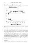
Purchased for the exclusive use of nofirst nolast (unknown)
From: SCC Media Library & Resource Center (library.scconline.org)