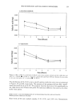
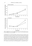
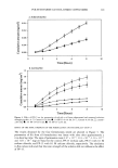
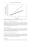
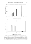
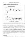
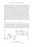
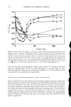
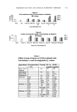
162 JOURNAL OF COSMETIC SCIENCE diameter (D) is the same as previously determined by karyometry. The corrected nucleus/ cytoplasm ratio will then be: Vvc Corrected n/c - -- 1 - Vvc where 1 - Vvc is the corrected volumetric cytoplasmic fraction (Vvcyt). Numerical nuclear density (Nvn). The area of the epithelium within the test system was evaluated by counting the points that fall on it, and the epithelial volume was propor- tional to it. The nuclei inside the standard square were then counted. The total area of the square was 50.625 pm 2 in two fields per section, for a total of 20 fields per block, and this permitted us to obtain the number of nuclear sections of the area (Nav). The number of nuclei per unit volume (numerical nuclear density, Nvn) was calculated using the Abercrombie (7) correlation modified by Elias eta/. (8): Sav Svn - D+T where D is the mean nuclear diameter previously estimated by karyometry, and t is the thickness of the section (6 pm). The result obtained corresponds to the number of nuclei per mm 3. External surface/basal layer (V/S) ratio. To determine this ratio we counted the number of times the test line intersected the interface under study (keratin or connective tissue). The V/S ratio is given by the equation: P'I V/S - 4I where P is the number of points that fall on the epithelium, I the number of intersections of the test line with the interface under study, and I the length of the test line, determined by the ratio: d'l I- 2 where d is the distance between two contiguous points marked on the test line. The fact that the epithelial volume (Vep) is constant for each field permits the estab- lishment of a direct relation between the surface areas of the two interfaces corresponding to the same standard volume: IK/Vep IK -- Ict/Vep Ict where IK and Ict are the numbers of intersections of the test line with the epithelium- keratin and epithelium-connective tissue intersections. The V/S ratio was inverted to obtain the IK/Ict ratio instead of the Ict/IK ratio. Cytoplasmic volume and epithelial cell volume. Cytoplasmic volume (Vct) was estimated from the previously determined nuclear volume and the corrected nucleus/cytoplasm ratio. In turn, the sum of the mean nuclear and cytoplasmic volumes provides the estimated value of the epithelial cell. The cytoplasmic volume is given by the ratio:

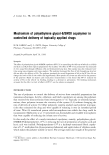
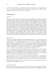
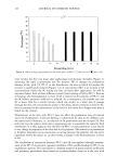
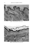
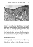
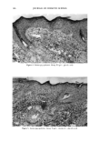
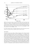
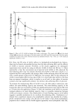
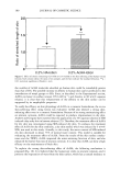
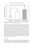
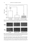
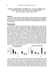
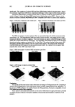
VITAMIN A AND GLYCOLIC ACID FORMULATIONS 163 Vct z Vn corrected n/c The volume of the epithelial cell, in turn, is given by the equation: Vcel = Vn + Vct Mean epithelial thickness. Mean epithelial thickness was estimated by the formula of Weibel (9): P-L 2(IK + Ict) where P is the number of points that fell on the epithelium, L is the length of the test line, and IK and Ict are the numbers of intersections of the test line with the epithelium- keratin interface and the epithelium-connective tissue interface, respectively. Statistical analysis. Data were analyzed statistically using nonparametric tests such as the Mann-Whimey and median tests. Data analysis and the mathematical calculations in- volved in the stereologic studies were performed using several programs elaborated in the Department of Stomatology, Dental School of Ribeirgo Preto, USP, by Profs. Geraldo Maia Campos and Miguel Angel Sala Di Matteo using ADVANCED BASIC language. RESULTS AND DISCUSSION HISTOPATHOLOGY Group I (control). The lining epithelium of the guinea pig epidermis is of the keratinized stratified pavement type (Figure 1). The basal layer is clearly visible, resting on the basement membrane and consisting of low cells with scarce cytoplasm and an ovoid nucleus slightly more stained than the nuclei of more superficial layers. The cells of this layer are well organized and arranged in such a way that the long axis is perpendicular to the basement membrane. The spinous layer, located above the basal layer, consists of more voluminous cells with nuclei containing sparse chromatin and clearly visible nucleoli. These cells tend to be arranged in such a way that the long axis is parallel to the surface. Above this layer is the granulose layer, whose cells contain keratin-hyaline granules in the cytoplasm. The horny layer is located in the outermost portion and consists of keratin filaments firmly adhering to the granulose layer. The dermis, located immediately below the epidermis, consists of a layer of connective tissue. Group II (gd on/y). In this group the epidermis was thicker, with barely developed papillae. The cells of the basal and spinous layers were more voluminous and the nuclei of weak chromatin were also more voluminous. Some of these cells were edematous. The granulose layer was clearly visible and the horny layer was thinner (Figure 2). Group III (gd + vitamin A). In this group, the aspect of the epidermis was quite similar to that observed in Group II. The cells of the basal and spinous layers, as well as their nuclei, were more voluminous. The thickness of the epithelium was more evident, and the basal and spinous layers were also thicker. The granulose layer presented cells filled
Purchased for the exclusive use of nofirst nolast (unknown)
From: SCC Media Library & Resource Center (library.scconline.org)