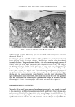
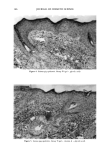
160 JOURNAL OF COSMETIC SCIENCE Vitamin A palmitate acts on the skin by keeping it in good condition and favoring its correct metabolism. It acts on epithelization in dry and rough skin, as well as on keratinization considered to be abnormal. Vitamin A palmirate has been used in der- mocosmetic preparations in combination with glycolic acid because the latter reduces the cohesion of corneocytes, stimulating skin desquamation, which in turn facilitates vitamin A absorption by the skin and leads to the expected results. Alpha-hydroxy acids are being incorporated into a new generation of treatment cosmet- ics. This is a new and interesting chapter in the formulation and scientific investigation of these products, since satisfactory results in the recovery of aged skin can be obtained with much simpler molecules. The objective of the present study was to investigate the histopathological alterations caused by dermocosmetic formulations containing vitamin A and/or glycolic acid in guinea pig skin, determined by appropriate stereologic techniques. EXPERIMENTAL PROCEDURE FORMULATIONS STUDIED We used a non-ionic gel formulation consisting of 2% hydroxy-ethyl-cellulose, 2% glycerin, 3% propyleneglycol, 0.2% methyldibromo-glutaronitrile and phenoxyethanol, 0.01% alpha-tocopherol and distilled water, with and without the addition of 0.5% vitamin A palmirate (1,000,000 IU/g) and/or 4.2% glycolic acid. BIOLOGICAL ASSAY Artfinals. Adult guinea pigs weighing on average 350 g were used. The animals were kept in individual cages and received commercial ration and green food (rami), as well as water ad/RSftum. Treatment. Five areas measuring 1 cm in diameter were shaved on each side of the dorsum of each animal, one of them used as control and the other four for the application of the cosmetic formulations. The formulations were applied daily for one week. The treatment was as follows: a) area I, no treatment (control) b) area II, application of the gel only c) area III, application of the gel with vitamin A added d) area IV, application of the gel with glycolic acid added e) area V, application of the gel with vitamin A and glycolic acid added. Histology. After one week of treatment the guinea pigs were sacrificed and skin fragments were obtained from each shaved area and immediately immersed in a fixing solution of 85 ml of 80% alcohol, 10 ml formalin, and 5 ml acetic acid. The fragments were fixed for 24 hours and then dehydrated, cleared, and embedded in paraffin. Serial 6-pm-thick sections were then obtained, and ten sections per block were obtained from a total of 500 sections, so that each of these ten sections would correspond to an interval of 50 sections. The sections were stained with hematoxylin and eosin. 3•orphometry and/•aryometry. For the morphometric study (analysis of the nucleus of the epithelial layers), the skin sections obtained from each experimental group were analyzed with a Henamed light microscope equipped with a 100x immersion objective and a light camera (Jena).

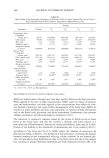
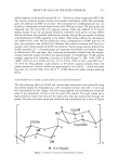
VITAMIN A AND GLYCOLIC ACID FORMULATIONS 161 The largest and smallest diameters of the nuclei of the basal and spinous layers of the epidermis were measured in drawings of the image projected onto paper at a final magnification of 1000x. The nuclear images obtained were traced with a no. 2 black pencil, with care taken to consider only elliptical images. The largest and smallest axes of these images were then measured with the aid of draft paper. The following karyomet- ric parameters were estimated: ß Mean diameter: M -- (D ß d) •/2 ß Perimeter: P -- (q'r/2) ß [1.5 ß (D + d) - M] ß Largest diameter/smallest diameter ratio: D/d ß Volume:V = 6 -• ß •'M 3 ß Area: A = q-r'M 2/4 ß Area/volume ratio: 3/2 ß M ß Shape coefficient: F = 4 ß q-r 'A/p2 ß Contour index: I = P/(A) •/• ß (D - d)•/•/D Stereo/ogy. In the present study we used a grid, idealized by Merz (2), printed on paper to draw the epithelial structures. The grid consists of a square that limits the test area, containing a system of points marked on a sinuous line formed by the succession of enchained semicircles. The Merz grid can be used to count points on a given histological structure, and also to count intersections between two contiguous structures, by con- sidering the number of points that fall on the structure under study in the former case and the number of times that neighboring surfaces cut the curved line in the latter. Thus, in order to obtain the nucleus-cytoplasm ratio, the thickness, the numerical nuclear density, the epithelial volume/interface ratio, the cytoplasmic volume, and the epithelial volume, we used point counting (2000 per animal, corresponding to the product of 20 microscope fields per 100 points on the grid) or the number of intersec- tions, according to the requirements of the stereologic equation with respect to the parameter studied. Nucleus/cytoplasm (n/c) ratio. The nucleus/cytoplasm ratio is given by the ratio of the relative volumes of nucleus and cytoplasm: Vvn n/c- Vvcyt The relative volumes are determined by the number of points falling on the structure considered (3-5). The value thus obtained is an overestimate of the real value due to the so-called "Holmes effect" (5), which results from the use of histological sections of finite thickness. To correct this overestimate it is necessary to take into account the size of the structure involved and the thickness of the histological section. Henning (6) proposed the following corrective formula for the Holmes effect, in which the nuclei are seen as if they were spheres of mean diameter D, and T is the thickness of the section: Vvn Vvc -- 1 + 3T/2D In this expression Vvc is the corrected volumetric fraction of the nuclei, and Vvn is the observed volumetric fraction calculated by dividing the number of points falling on the nuclei by the total number of points falling on the nucleus and cytoplasm. The mean
Purchased for the exclusive use of nofirst nolast (unknown)
From: SCC Media Library & Resource Center (library.scconline.org)