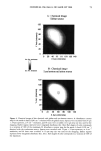
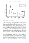
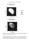
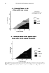
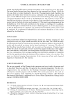
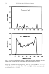
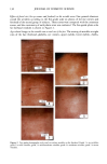
j. Cosmet. Sci., 51, 73-90 (March/April 2000) Chemical imaging of hair by infrared microspectroscopy using synchrotron radiation JEAN-LOUIS BANTIGNIES, G. L. CARR, DOMINIQUE LUTZ, SYLVIE MARULL, GWYN P. WILLIAMS, and GILBERT FUCHS, The Elf Atochem Company, CRRA, Rue Henri Moissan, BP 63, 69310 Pierre Bgnite, France (J-L.B., G.F.), National Synchrotron Light Source, Brookhaven National Laboratory, Upton, NY 11973 (G.L.C., G.P.W.), and The Yves Rocher Company, 101 Quai du Prgsident Roosevelt, 92444 Issy-les-Moulineaux Cedex, France (D. L., S.M.). Accepted for publication February 15, 2000. Synopsis For the first time, infrared microspectroscopy with a synchrotron source has been used to characterize human hair before and after bleaching. This high-brightness source allows a lateral resolution approaching the diffraction limit (a few microns) to be achieved. Thus, 4-micron diameter areas have been analyzed, and chemical imaging of hair cross sections obtained with good contrast. In particular, the variations of the integrated absorbances of the amide stretching vibration (3290 cm -•) and CH stretching vibrations (be- tween 3000 and 2800 cm -•) as a function of sample position have allowed chemical imaging of the medulla and cuticle. We also studied bleaching of the hair, and we checked different chemical effects as a function of position in the hair cross section. We also observed a non-homogeneous distribution of bonded water. The trans- formation, due to breakage of a specific chemical group, was observed and spatially characterized. The sulphonate contributions were given special attention and showed non-homogeneous distributions within the hair. INTRODUCTION Using infrared microspectroscopy (IMS) with synchrotron radiation, the cuticle, cortex, and medulla can be differentiated within virgin hair samples. Human hair has been studied previously by infrared spectroscopy, and the various characteristic absorption frequencies of the predominant keratin protein are well established (1-3). Such inves- tigations were carried out using diffuse or attenuated total reflectance for which sample preparation methods can lead to problems of reproducibility (4-6). More definitive Address correspondence to Sylvie Marull. 73

74 JOURNAL OF COSMETIC SCIENCE information could be obtained by studying intact individual hair fibers, but the small diameter of the hair fibers makes such measurements difficult (100 pm). The problems associated with the small sample dimensions have been overcome (7,8) by combining the chemical specificity afforded by infrared spectroscopy with the lateral resolution of IMS. Indeed, using a double-aperture experimental arrangement to reduce spurious signals from neighboring areas (9), regions down to =30 microns in size have been analyzed (10). Though individual hair shafts are small, their thickness still exceeds the optical penetration depth at wavelengths where absorption is quite strong. By means of IMS, flattened hair shafts treated with oxidizing agents have been examined at different distances from the root to the tip (6). Using a diamond "squeeze-cell" to effectively thin the hair, characterization by IMS of hair samples from the anagen to the telogen phases has also been made by probing the fiber from the bulb to the shaft (11) while the sensitivity of cortical hair cells in contact with potassium hydroxide in 1-butanol solutions has been tested using IMS (12). IMS has also been employed to examine the inner part of a microtomed hair to determine drug ingestion (13-15), and absorbance contour linear maps have been obtained. Nevertheless, IMS experiments using a thermal source (globar) are limited, by the brightness of the source, to a lateral resolution above 24 x 24 pm (5,10). Replacing the traditional thermal source of a conventional IR microscope with a high-brightness synchrotron source allows substantial improvements of both signal-to-noise ratio and lateral resolution of IMS measurements (16,17). Thus, experiments can be carried out in diffraction-limited conditions, and an image of a hair sample can be obtained with a lateral resolution of few microns. IMS with synchrotron radiation was recently performed to study the localization of drug metabolites within longitudinally microtomed sections of human hair (15). In this investigation, a synchrotron IMS study of hair shaft samples was performed. We present for the first time--detailed, high contrast, chemical images of transverse hair cross sections. In particular, the cortex, cuticle, and medulla are differentiated. Results are compared with those obtained using a standard thermal source. Bleached hair was also imaged chemically with a lateral resolution of a few microns. MATERIALS AND METHODS SAMPLE PREPARATION Untreated, dark homogeneous human hair fibers of European origin were used. Tresses approximately 15 cm in length were first separated into 2-g pieces. A bleaching cream was prepared by blending one volume of commercial bleaching powder (Platifiz © from l'Oreal Inc.) with two volumes of hydrogen peroxide solution (40 volume). To complete the bleaching process, bleaching cream was uniformly applied on dry hairpieces (10 g per 2-g piece). Samples were then wrapped in an aluminum sheet and left at ambient temperature for one hour. The hair samples were then washed with water and retreated with the bleaching cream for another hour. Finally, the hair samples were water rinsed and allowed to dry at ambient temperature. SAMPLING FOR IMS EXPERIMENTS For the IMS experiments, the hair fibers were first cut into 1-cm lengths starting
Purchased for the exclusive use of nofirst nolast (unknown)
From: SCC Media Library & Resource Center (library.scconline.org)