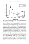
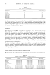
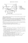
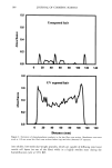
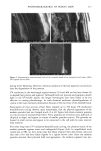
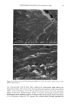
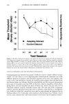
104 JOURNAL OF COSMETIC SCIENCE ponent of sunlight is known to be responsible for these changes. In recent years, UV radiation at lower wavelengths has significantly increased due to the deterioration of the ozone layer. While the earth's atmosphere filters out most radiation below 295 nm, depletion of the ozone layer, and therefore the reduction in the screening effect of the atmosphere, permits lower wavelength components to reach the earth's surface. These lower wavelength regions of the UV radiation received by the earth are the most energetic and therefore can cause severe photodegradation. The UV range of the sunlight can be divided into three wavelength regions, namely UV-A (320-400 nm), UV-B (280-320 nm), and UV-C (280 nm). UV-C radiation is totally filtered out by the atmosphere and is only experienced in space. We have carried out extensive studies, investigating by various microscopic techniques the effects of photochemical oxidation on specific aspects of hair damage such as the appearance of the surface cuticle cell, intercellular cohesion, scale lifting during longi- tudinal extension, the integrity of the cuticular sheath, the physical nature of the melanin granules, characteristic fracture patterns, and loss in hair color. In other words, we have examined radiation-induced changes in the physical rather than chemical nature of the hair fiber. Hair fibers used in these studies were exposed to UV radiation/ humidification cycling in a QUV Accelerated Weathering Tester. Various follow-up treatments of these long-term UV-irradiated fibers illustrate the extent of photodegra- dation inflicted upon the hair proteins. More recently, we have also examined the effects of both the relative humidity and the spectral energy distribution in the radiation on the photochemical oxidation of the hair fiber. Comparisons were made between the results obtained at various relative humidi- ties in two different fading units, namely, the QUV Accelerated Weathering Tester (290-400 nm UV-A kn• = 340 nm) and the Atlas Weather-Ometer ©, "AW" (Xenon solar simulator, 250-800 nm). Using FESEM, we characterized by different approaches the extent of photodamage inflicted upon the physical nature of the hair fiber in general and the cuticula in particular. EXPERIMENTAL MATERIALS Hair type. Root sections of 14-in-long, brown European hair fibers from DeMeo Brothers were used. The major axis of these hair fibers ranged from 70 lam to 120 lam. Because of this variation in size, single hair fibers also varied in the depth of shade. Fibers with the larger major axis were more elliptical than fibers with a smaller major axis. UV EXPOSURE CONDITIONS QUV Accelerated Weathering Tester. The QUV simulates the sunlight in the range of 290-400 nm, with an irradiance maximum of the fluorescent bulb at 340 nm. The irradiance intensity factor has been chosen to be 1.35 compared to 1.0 for regular sunlight. The energy density at the 340-nm wavelength is kept constant at 0.97 W/m 2. The total energy density of the UV light in the wavelength range of 300-400 nm is 5.06 mW/cm 2. Hair fibers were exposed to two sets of conditions in the QUV: (a) 0, 100, 300, 500, and

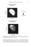
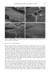
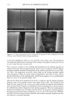
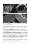
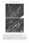
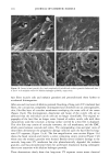
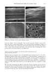
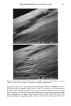
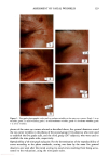
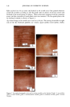
PHOTODEGRADATION OF HUMAN HAIR 105 700 hours at alternating three-hour cycles of humidification (at 95 % RH 40øC) and UV irradiation (50øC RH decreasing from 95% to 10% within 30 min, then remaining at 10% for 2.5 h) and (b) 0, 100, 200, and 300 hours continuous UV irradiation at constant 10% RH and 50øC. At/as Weather-Ometer ©, ("AW"). The "AW" has a spectral distribution ranging from UV through the visible range, simulating sunlight. These exposure conditions correspond to an "average" 45 ø Miami summer sunlight. The energy density at the 340-nm wave- length is kept constant at 0.3 W/m i . This means that at the specific wavelength of 340 nm, the energy density in the QUV (0.97 W/m 2) is approximately three times greater than that of the "AW" (0.3 W/mP). However, in the wavelength range of 300-400 nm, the energy densities of the QUV (300-400 nm yield 5.06 mW/cm 2) and the "AW" (300-400 nm yield 4.46 mW/cm 2) are similar, although the distribution is quite different. The specific spectral distributions in the "AW" in the range of 250-300 nm yield 0.012 mW/cm •, and 400-800 nm yield 36.80 mW/cm 2. The total energy density in the "AW" is 41.272 mW/cm •. Specific exposure conditions of the hair fibers to UV/visible radiation in the "AW" were 0, 100, 200, and 300 hours at constant 20%, 50%, and 70% RH and 50øC. Post-treatment of hair fibers in alkaline hydrogen peroxide. Hair fibers exposed to UV irra- diation/humidification (at 95% RH) cycling were subjected to post-treatment in 6% alkaline hydrogen peroxide from seconds up to two hours. This was done to illustrate and characterize the severe extent of photodegradation inflicted upon the fiber during treat- ment in the QUV. Post-treatment of samples irradiated at low and intermediate humidities with water. UV irra- diation/humidification (at 95% RH) cycling lead to extensive "thinning" of the surface cuticle cells and "fusion" of the scale edges. Differentiation of the surface cuticle cell, so characteristic of the untreated hair fiber, eventually disappears at longer exposure times. We concluded that the presence of moisture in the highly swollen fiber during the humidification cycle is responsible for transporting the degraded, low-molecular-weight protein fragments out of the cuticle cell and possibly into the cortex, thereby causing the collapse and thinning of the surface cuticle cell. Hair fibers irradiated at low humidities (from 10% to 70% RH) in the two fading units do not show this feature of thinning and fusion of the cuticle cell. Apparently, the lack of mobile water in the hair fiber and the lack of swelling of the fiber eliminate the transport of photodegraded fragments out of the cuticle cell. To mobilize the photode- graded proteins, hair fibers irradiated under low RH conditions in both the QUV and "AW" were subjected to a water post-treatment. The fibers were immersed for 60 minutes in lukewarm, deionized water, air-dried, and then examined longitudinally in the SEM. This was to establish whether post-treatment in warm water would result in thinning and fusion of the cuticle cell by diffusing photodegraded materials out of the cuticle cell, assuming that photo-oxidation of the hair proteins had occurred at all. INVESTIGATIVE METHOD Field emission scanning electron microscopy. Longitudinal and cross-sectional segments of untreated and UV-exposed fibers were mounted on double-sided tape and coated with approximately 90 fk of platinum. The hair fiber topography and interior were examined
Purchased for the exclusive use of nofirst nolast (unknown)
From: SCC Media Library & Resource Center (library.scconline.org)