j. Cosmet. Sci., 51,367-377 (November/December 2000) Characterization of the physical factors affecting nail permeation using water as a probe GOURI GUPCHUP MALHOTRA and JOEL L. ZATZ, Unilever Research U.S., 45 River Road, Edgewater, NJ 07020 (G.G.M.), and College of Pharmacy, Rutgers University, 160 Frelinghuysen Road, Piscataway, NJ 08854 (J.L.Z.). Accepted for publication September 14, 2000. Poster entitled "Methodology for Studying Nail Permation In Vitro" presented at the Annual Scientific Seminar of the Society of Cosmetic Chemists, Chicago, May 1999. Synopsis Our research objectives were to (a) develop a reproducible technique to measure nail permeation in vitro, (b) study variations in nail permeability as a function of location (toenails vs fingernails), and (c) study the effect of pH and temperature. We monitored the permeation of tritiated water from aqueous gels using specialized Franz-type diffusion cells. For pH studies, the gels were buffered at pH 2, 7, and 12. For temperature studies, three settings (37øC, 47øC, and 57øC) were investigated. Our results showed that there was a large interindividual variability in nail permeation. Normalization of the data for toenail thickness reduced the standard deviation for flux from 42% to 21% of the mean value. Repeated measurements resulted in less than 5% variability, indicating that the technique was reproducible. Thumbnails, despite their reduced thickness, resulted in water fluxes equal to those of toenails, suggesting the presence of a tighter keratin network. Although not conclusive, our results suggest that thumbnails also differ in structure from other fingernails. While pH did not affect permeation through nails, increased temperature significantly enhanced water permeation. The effects of pH and heat treatment were reversible. INTRODUCTION The permeability properties of the human nail were summarized in a recent review (1). It is now generally accepted that the human nail is primarily composed of a highly cross-linked keratin matrix that gives the nail its effective barrier properties. The nail keratin itself is composed of or-helical filaments and non-helical matrix keratins that are rich in the sulfur-containing amino acid half-cystine. This results in numerous disulfide linkages throughout the matrix that give rise to its extensively cross-linked structure and resistance to penetration (2,3). The nail has been documented to be more permeable to small, hydrophilic molecules such as water, methanol, and ethanol, than to larger, lipophilic molecules (4,5). Some attempts have been made to characterize the effect of pH by using ionizable drugs however, it is not clear whether pH alters the nature of the nail plate itself or whether better permeation under certain pH conditions results from 367

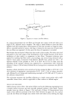
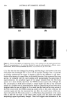
368 JOURNAL OF COSMETIC SCIENCE the state of dissociation of the drug (5-7). To our knowledge, there are no reports in the literature on variations in nail permeability as a function of different sources of nails. The effect of environmental factors such as pH and temperature on intrinsic nail permeability properties also remains to be investigated. The objectives of our research were to (a) develop a reproducible technique to measure nail permeation in vitro, (b) study variations in nail permeability as a function of differing source (between donors, toenails vs fingernails), and (c) study the effect of pH and temperature on nail permeation. In our experiments, tritiated water was chosen as a marker molecule, and its permeation through nails was monitored as a function of various environmental and physical factors. Water was expected to be non-destructive to the nail and is a good marker because of its small size. It was first necessary to develop a suitable technique to reproducibly measure nail permeation in vitro. For this purpose, specialized diffusion cells that had been adapted to hold the human nail were used, and water permeation through the nails was repeatedly monitored. EXPERIMENTAL MATERIALS Tritiated water (3H20) (specific activity of 1 l•Ci/mg) was obtained from NEN TM Life Sciences Products (Boston, MA). Hydroxyethyl cellulose (HEC, Natrosol ©, Aqualon Co., Wilmington, DE) and polyethylene glycol-20-oleyl ether (PEG-20-oleyl ether, Croda, Inc., Parsippany, NJ) were used as received. All other laboratory chemicals (ACS grade or better), including Scintiverse I, were obtained from Fisher Scientific (Springfield, NJ) and used as received. Cadaver toenails or fingernails were obtained (frozen) from tissue banks [International Institute for the Advancement of Medicine (IIAM), Scranton, PA Advanced Biosurfaces Inc., Minnetonka, MN]. DEVELOPMENT OF METHODOLOGY FOR IN VITRO NAIL PERMEATION STUDIES Preparation of nails. Human toenails/fingernails were obtained (frozen) from tissue banks based on a protocol for size, patient history, etc. Of all the toenails, only the great toenail was large enough to fit in the diffusion assembly however, all the fingernails (thumbnail and other fingernails) were suitable for use. The nails were thawed at room temperature for one hour, and the adhering skin and tissue were removed with a pair of scissors and a scalpel. The nails were cleaned by rinsing them in a mild detergent solution (1%), followed by two rinses in aleionized water (DI water). The thickness of the nails was measured with a micrometer, and the nails were immersed in 10 ml of DI water for 24 hours to allow complete hydration just prior to use in permeation experiments. Nail permeation cells. Franz-type diffusion cells (Crown Glass Co., Somerville, NJ), spe- cially designed to hold the human nail were used (Figure 1). In this cell, the donor compartment (Teflon) had a two-part construction to hold the nail. The lower half of the donor compartment screwed on to the glass receptor chamber, the nail was placed above this, and the upper half of the donor compartment was placed above the nail. The two parts of the donor compartment were clamped together to hold the nail in place between
Purchased for the exclusive use of nofirst nolast (unknown)
From: SCC Media Library & Resource Center (library.scconline.org)