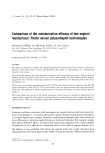
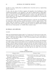
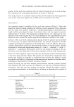
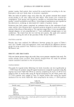
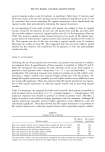
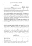
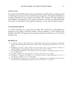
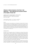
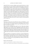
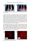
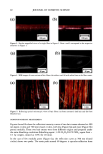
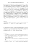
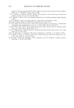
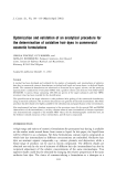
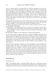
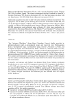
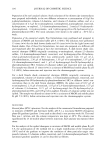
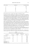
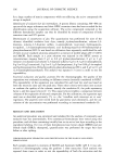
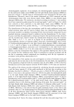
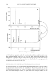
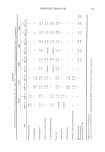
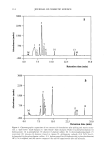
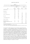
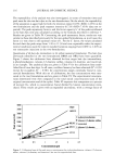
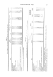
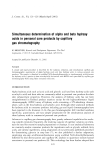
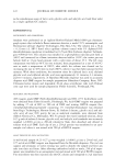
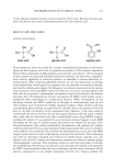
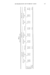
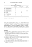
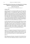
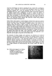
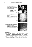
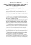
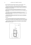
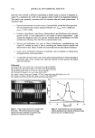
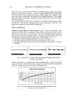
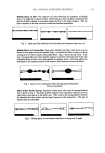
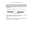
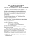
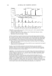
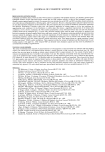
MEDULLA STRUCTURE AND HAIR APPEARANCE 91 (a) (b) ß ß ß ß I i ß 10 cm 10 cm Figure 1. Images for hair tresses evaluated by selected panelists as (a) shiny and brilliant and (b) dull and whitish in appearance. (la), and dull and whitish (lb) in appearance, respectively. There was little difference found when cuticle surfaces were observed using SEM. Figures 2a and 2b show optical microscopic views of the hair bundles of Figures la and lb, respectively, observed by a reflecting optical microscope with a low magnification. White lines are obvious in the whole view of Figure 2b. Further magnified views of a single hair fiber are shown in Figures 3a and 3b, corresponding to Figures la and lb, respectively. White lines are located at the fiber center of Figure 3b, whereas no such lines are observed in Figure 3a, implying that the differences in the appearance originated from the state of the medulla. Figures 4a and 4b show the SEM views of cross sections of the fiber without and with, respectively, the white lines shown in Figures 2 and 3. It was confirmed that the white lines are actually identical with light scattering at the microporous structure of the medulla and thus significantly influence hair appearance. Light penetrating deep into the center of the fiber is scattered at porous medulla, and the scattered light reaching the surface of the fiber is perceived by the observer. There- fore, the influence of porous medulla on hair appearance is greater in bleached or colored hair than in dark hair. Figure 5 shows the differences in the microscopic views of tip parts of dark hair and bleached hair obtained from the same origin, where the amount of porous medulla is confirmed to be comparable. In the case of dark hair (Figure 5a), only split ends are observed as whitish, while in bleached hair not only split ends but also porous medulla are observed as whitish (Figure 5b). (a) . (b) I mm 1 nun Figure 2. Reflecting optical microscopic views of hair fibers: fibers from hair evaluated by experts as (a) shiny and brilliant and (b) white, chalky, and lusterless.

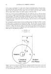
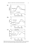
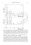
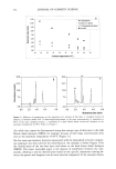
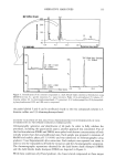
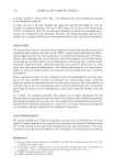
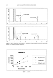
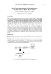
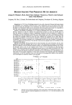
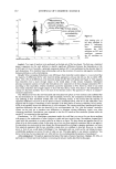
92 JOURNAL OF COSMETIC SCIENCE (a) (b) ß ! ! i I I 1 O0 Ixm 100 pm Figure 3. Further magnified views of a single fiber in Figure 2. Parts a and b correspond to the respective notations in Figure 2. (a) (b) : i : 1 50 I•m 50 I•m Figure 4. SEM images of cross sections of hair fibers: (a) without and (b) with white lines at the fiber center (a) ' (b) ' ! ! [ I mm I mm Figure 5. Reflecting optical microscopic views of hair fibers: (a) from untreated dark hair and (b) from bleached hair. GONIOPHOTOMETRIC MEASUREMENT Figures 6a and 6b show the reflection intensity curves of two hair tresses observed at 400 nm (open circles) and 700 nm (closed circles), with less (Figure 6a) and more (Figure 6b) porous medulla. These two hair tresses were from different origins and prepared under the same bleaching conditions (bleaching agent: 3.4% H202/0.67% NH3 agent/hair = 1/1 by weight, treated at 30øC for 20 min). In the case of few medulla pores (Figure 6a), the reflection curve at 700 nm (closed circles) shows two peaks. The main peak around 40 degrees is specular reflection from
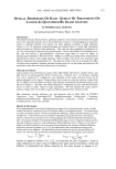
Purchased for the exclusive use of nofirst nolast (unknown)
From: SCC Media Library & Resource Center (library.scconline.org)