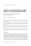
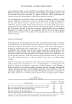
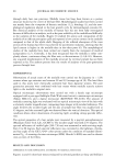
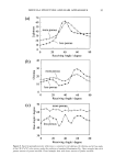
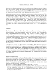
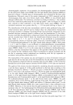
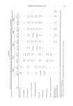
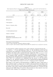
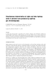
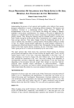
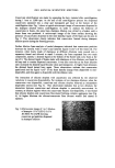
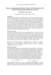
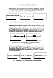
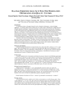
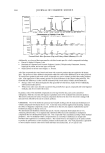
2001 ANNUAL SCIENTIFIC MEETING 145 THE DIFFERENTIAL DISTRIBUTION PATTERNS OF TRANSFERRED MELANOSOMES THE KERATINOCYTE IS REGULATED BY TuE RECIPIENT Id•ERATINOCYTE Ljiljana Minwalla, Ph.D.', Yang Zhao', I. Caroline LePoole, Ph.D. a, R.Randall Wickett, Ph.D? and Raymond E. Boissy, Ph.D.' 'University of CincinnatL Department of Dermatology, Cincinnati, OH •Loyola University of Chicago, Department of Pathology, Maywood, IL and sUniversity of CincinnatL Department of Pharmaceutical Sciences, CincinnatL OH Melanocytes are the cells in the body that are responsible for pigmentation. Melanocytes produce pigment within specialized organelles called melanosomes. In the skin, melanocytes transfer these pigment-filled melanosomes to surrounding keratinocytes. Without this transfer process, pigmentation does not occur. The melanosomes that are found in keratinocytes of Black skin are larger and distributed individually whereas those within keratinocytes of Caucasian skin are smaller and distributed in clusters. This is an important feature that lends itself to differences in complexion coloration. When melanosomes are larger and more spread out, they can absorb more incoming light so that less is refracted. This gives skin a darker coloration. When the melanosomes are smaller and distributed in clusters, they absorb less light and more is refracted thus giving skin a lighter coloration. Figure 1 exhibits the typical differences in melanosome distribution panems that exist between Black and Caucasian skin. Melanosomes within Black keratinocytes (Figure 1 A) are distributed individually throughout the cytoplasm, being predominantly localized apically over the nucleus. In contrast, melanosomes within Caucasian keratinocytes (Figure 1 B) are almost exclusively membrane-bound in clusters but also predominantly localized over the nucleus. Figure I Comparable differences between the distribution ofmelanosomes by histological examination of Black and Caucasian skin. Melanosomes in Black skin (A) are singly distributed throughout the epidermal keratinocytes. Melanosomes in Caucasian skin (B) are found as membrane- bound clusters. (Illustration courtesy of Dr. Raymond E. Boissy).

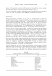
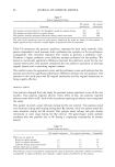
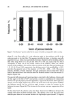
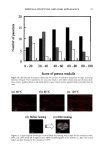
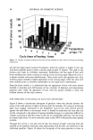
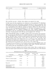
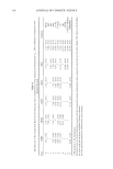
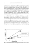
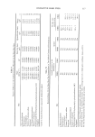
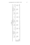
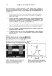
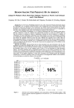
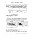
146 JOURNAL OF COSMETIC SCIENCE The disparity in distribution patterns of melanosomes is one factor that gives differences in skin pigmentation and in photoprotection. The control of these innate distribution patterns of melanosomes is poorly understood. To investigate this process, cocultures ofmelanocytes and keratinocytes from different racial backgrounds were examined using electron microscopy. Melanosomes in keratinocytes were counted and categorized as individual or in clusters of 2-3, 4-6, or greater than 6. In addition, melanosomal area wa•, determined for individually versus clustered melanosomes to correlate with the different distribution patterns observed. Results indicate that in our model system, melanosomes in keratinocytes from different racial backgrounds have a combination of clustered and individual melanosomes. When dark skin derived keratinocytes were cocultured with melanocytes derived from (a) dark or (b) light skin, recipient melanosomes were individual versus clustered in (a) 77.6% versus 22.4% and (b) 63.5% versus 36.5%, respectively. In contrast, when light skin derived keratinocytes were cocultured with melanocytes derived from (c) dark or (d) light skin, recipient melanosomes were individual versus clustered in (c) 33.5% versus 66.5% and (d) 38.7% and 61.3%, respectively. These results indicate that regardless of the donor melanocyte, recipient melanosomes will be distributed by keratinocytes from dark skin predominantly individually, and from light skin predominantly in membrane-bound clusters. One factor that appears to regulate the distribution patterns of melanosomes is their size. Studies regarding this are in dispute. Therefore, we assessed our cocultures for the pattern ofmelanosome distribution within keratinocytes and in turn correlated them with cell donor type and melanosome size. We found that although there were differences in melanosome size from dark or light donor melanocytes, there were no dift•rences in the sizes of melanosomes distributed individually compared to those clustered. This is demonstrated in Table 1. Table I Sizes of Melanosomes Donated either from Black of Caucasian Melanocytes within Keratinocytes. Sizes of Individually Distributed Melanosomes can be Compared with Sizes of Clustered Melanosomes. ire Average Melanosome Size (F.m 2) )m Melanosomes tual Melanosomes red Melanosomes •s + Caucasian ( 102ñ8 14X 10 • X 102+_6.71 X 10 • X 10•_+6 66 X 10 • __ __ locytes + Caucasian X 10-• ñ 2.89 X 10 '• X 10'2_+2 91 X 10 -• X 10'•_+ 4 06X 10 '• These results suggest that the size of the recipient melanosome does not appear to regulate how melanosomes will be distributed but instead some regulatory factor within the keratinocyte coordinates this process.
Purchased for the exclusive use of nofirst nolast (unknown)
From: SCC Media Library & Resource Center (library.scconline.org)