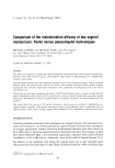
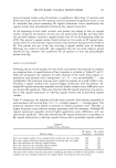
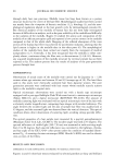
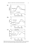
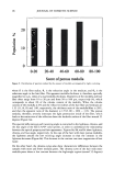
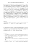
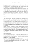
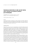
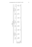
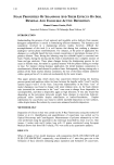
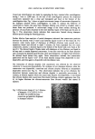
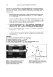
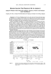
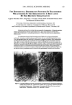
90 JOURNAL OF COSMETIC SCIENCE through daily hair care processes. Medulla tissue has long been known as a porous structure located at the center of the hair fiber. Morphological studies have been carried out mainly from the viewpoint of forensic medicine (1,2), histology (3), and the mor- phological regulation related to the hair growth cycle by dermatological concerns (4). But chemical analysis of the medulla of human hair has not been fully investigated because of difficulties in analysis, such as the poor solubility of the medulla and difficulty in the isolation of the medulla. Rogers (5) studied the amino acid composition of the medulla of an African porcupine quill and reported a low cystine content in the medulla compared to that of the whole shaft. Mapping of the infrared absorption of the cross section of the human hair fiber was achieved by synchrotron radiation, showing that the lipid content is higher in the medulla than in the other parts (6). The morphological studies of the medulla have been carried out mainly from the viewpoint of medical jurisprudence (2,7). Generally, it has been recognized that the medulla is either com- pletely absent, continuous along the fiber axis, or discontinuous (3-4,8-10). However, any acquired transformation of the medulla structure by external stimuli has not been reported so far. The authors present here the results of analyses of the pore-generation process through heat. EXPERIMENTAL Observations of actual states of the medulla were carried out for Japanese (n = 120) women whose age variation was between 20 and 49 (average age of 30). The hair fibers used in this study were chemically untreated hair from Japanese women. The pore generation processes were confirmed with hair tresses whose medulla scarcely scatters light in the medulla's original state. Optical microscopic observations were carried out with a handy type microscope equipped with a pen-type flashlight (Peak Wide-stand-micro), a stereoscopic microscope (Nikon SMZ-10), and a digital microscope (KEYENCE VH-6100). The amount of medulla scattering light was evaluated with an optical microscopic view of the hair with a relatively smaller magnification, comparing these images with standard references. The angle between the incident light and the axis of sample hair fiber was usually fixed at 45 degrees, and the scattered light was observed from the vertical direction. This optical condition allows direct observation of scattering light, avoiding strong specular reflec- tion. The optical properties of a hair sample were measured by a spectral goniophotometer (Murakami Color Tech. Lab., GCMS-3). The incident angle was fixed at 45 degrees. The spectral reflectance of visible light (390-730 nm) was measured at each receiving angle from 0 to 80 degrees. Each spectral reflectance was converted to the lightness, chroma, and hue angle of the CIE L*a*b* color system under the condition of standard illumi- nation D65. A scanning electron microscope (SEM, Hitachi S-4000) was used to observe cross sections of the fibers. RESULTS AND DISCUSSION DIFFERENCE IN HAIR APPEARANCE ACCORDING TO MEDULLA STRUCTURE Figures la and lb show hair tresses evaluated by selected panelists as shiny and brilliant

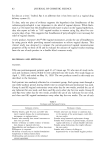
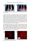
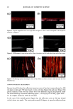
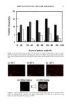
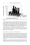
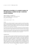
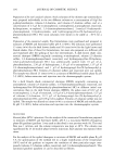
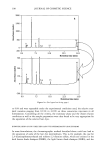
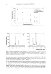
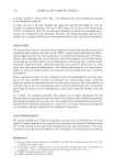
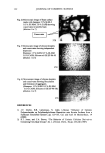
MEDULLA STRUCTURE AND HAIR APPEARANCE 91 (a) (b) ß ß ß ß I i ß 10 cm 10 cm Figure 1. Images for hair tresses evaluated by selected panelists as (a) shiny and brilliant and (b) dull and whitish in appearance. (la), and dull and whitish (lb) in appearance, respectively. There was little difference found when cuticle surfaces were observed using SEM. Figures 2a and 2b show optical microscopic views of the hair bundles of Figures la and lb, respectively, observed by a reflecting optical microscope with a low magnification. White lines are obvious in the whole view of Figure 2b. Further magnified views of a single hair fiber are shown in Figures 3a and 3b, corresponding to Figures la and lb, respectively. White lines are located at the fiber center of Figure 3b, whereas no such lines are observed in Figure 3a, implying that the differences in the appearance originated from the state of the medulla. Figures 4a and 4b show the SEM views of cross sections of the fiber without and with, respectively, the white lines shown in Figures 2 and 3. It was confirmed that the white lines are actually identical with light scattering at the microporous structure of the medulla and thus significantly influence hair appearance. Light penetrating deep into the center of the fiber is scattered at porous medulla, and the scattered light reaching the surface of the fiber is perceived by the observer. There- fore, the influence of porous medulla on hair appearance is greater in bleached or colored hair than in dark hair. Figure 5 shows the differences in the microscopic views of tip parts of dark hair and bleached hair obtained from the same origin, where the amount of porous medulla is confirmed to be comparable. In the case of dark hair (Figure 5a), only split ends are observed as whitish, while in bleached hair not only split ends but also porous medulla are observed as whitish (Figure 5b). (a) . (b) I mm 1 nun Figure 2. Reflecting optical microscopic views of hair fibers: fibers from hair evaluated by experts as (a) shiny and brilliant and (b) white, chalky, and lusterless.
Purchased for the exclusive use of nofirst nolast (unknown)
From: SCC Media Library & Resource Center (library.scconline.org)