145 Repairing Bleach-Damaged Hair
G, Japan Chlorophyll, Saitama, Japan), Fluorescein-4-isothiocyanate (FITC-I) (Dojindo,
Kumamoto, Japan), 2,2-Dithiobis(5-nitropyridine) (DTNP) (Sigma-Aldrich, Tokyo, Japan)
and 10–20 wt% polyacrylamide gradient gel plates (Kishida Chemical, Osaka, Japan)
were also commercially available. The bleaching was conducted using a mixture of Milbon
Powder Bleach® (Milbon, Tokyo, Japan) and Ordeve Oxidane® 6.0 (Milbon, Tokyo,
Japan) the former consisted of alkaline components and persulfate salt, and the latter
contained 6 wt% hydrogen peroxide as an essential ingredient.
PREPARATION OF HAIR SAMPLES
Bleaching treatment. The previously mentioned bleaching and oxidizing agents were mixed
in a ratio of 1:2 (w/w). After immersing the black hair in the aqueous mixture at a bath
ratio of 1:50 (w/w) at 25°C for 30 minutes, the fibers were washed well with water and dried
at 60°C with a hairdryer. These operations were repeated three times. The resulting hair
will be from now on be referred to as bleached hair.
Cross-linking treatment of hair (a general procedure). The bleached hair fibers were immersed in
pure water or a mixture of an aqueous solution of the polyphenol (8.4 mM) and copper (II)
chlorophyllin (4 ppm) at a bath ratio of 1:50 (w/w) at 25°C for 12 hours. The resulting hair
samples were rinsed with water and dried by using a hairdryer.
Cross-linking treatment of keratin proteins (a general procedure). The water-soluble keratin was
diluted with the phosphate buffer (PBS) (0.1 M, pH 7.4) to 5 wt%. Similarly, the PBS
solution of polyphenol (hydroquinone, catechol, resorcinol and chicoric acid) was prepared
at the appropriate concentration. The reaction was started by adding an aqueous solution
of copper (II) chlorophyllin (0.060mL) and the polyphenol solution (0.24mL) to the protein
solution (2.7mL). After standing still at 40°C for 12 hours, the reaction mixture was
obtained. In control experiments of the reaction, the keratin was replaced with an equal
volume of lysozyme (0.8 wt%).
SODIUM DODECYL SULFATE POLY(ACRYLAMIDE) GEL ELECTROPHORESIS (SDS-PAGE)
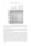
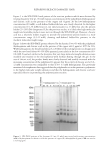
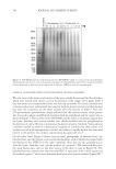
The SDS-PAGE was carried out in a manner similar to a previous method.13 The
polyphenol treated keratin solution (10 µL) was diluted with the X2 Laemmli buffer (Bio-
Rad Laboratories, Hercules, California, USA) (10 µL). Each of the mixtures was heated at
100°C for 5 minutes, cooled to ambient temperature and applied to the polyacrylamide
gradient gel plate (10-20 wt%). The resulting gel plate was subjected to electrophoresis
at 20 mA for about 90 minutes. The protein molecular weight maker II (TEFCO, Tokyo,
Japan) was also electrophoresed at the same time. The protein bands were visualized by
staining with Coomassie brilliant blue (CBB).
AMINO ACID ANALYSIS
The amino acid analyses of the polyphenol treated keratin and hair were performed
according to a high-speed RP-HPLC/FL method.14,15 Polyphenol treated product (1mg) was
hydrolyzed in the vapor phase with 6 M HCl at 110°C for 24 hours according to the PicoTag
protocol (Waters Corporation, Milford, Massachusetts, USA). The resulting hydrolysate
G, Japan Chlorophyll, Saitama, Japan), Fluorescein-4-isothiocyanate (FITC-I) (Dojindo,
Kumamoto, Japan), 2,2-Dithiobis(5-nitropyridine) (DTNP) (Sigma-Aldrich, Tokyo, Japan)
and 10–20 wt% polyacrylamide gradient gel plates (Kishida Chemical, Osaka, Japan)
were also commercially available. The bleaching was conducted using a mixture of Milbon
Powder Bleach® (Milbon, Tokyo, Japan) and Ordeve Oxidane® 6.0 (Milbon, Tokyo,
Japan) the former consisted of alkaline components and persulfate salt, and the latter
contained 6 wt% hydrogen peroxide as an essential ingredient.
PREPARATION OF HAIR SAMPLES
Bleaching treatment. The previously mentioned bleaching and oxidizing agents were mixed
in a ratio of 1:2 (w/w). After immersing the black hair in the aqueous mixture at a bath
ratio of 1:50 (w/w) at 25°C for 30 minutes, the fibers were washed well with water and dried
at 60°C with a hairdryer. These operations were repeated three times. The resulting hair
will be from now on be referred to as bleached hair.
Cross-linking treatment of hair (a general procedure). The bleached hair fibers were immersed in
pure water or a mixture of an aqueous solution of the polyphenol (8.4 mM) and copper (II)
chlorophyllin (4 ppm) at a bath ratio of 1:50 (w/w) at 25°C for 12 hours. The resulting hair
samples were rinsed with water and dried by using a hairdryer.
Cross-linking treatment of keratin proteins (a general procedure). The water-soluble keratin was
diluted with the phosphate buffer (PBS) (0.1 M, pH 7.4) to 5 wt%. Similarly, the PBS
solution of polyphenol (hydroquinone, catechol, resorcinol and chicoric acid) was prepared
at the appropriate concentration. The reaction was started by adding an aqueous solution
of copper (II) chlorophyllin (0.060mL) and the polyphenol solution (0.24mL) to the protein
solution (2.7mL). After standing still at 40°C for 12 hours, the reaction mixture was
obtained. In control experiments of the reaction, the keratin was replaced with an equal
volume of lysozyme (0.8 wt%).
SODIUM DODECYL SULFATE POLY(ACRYLAMIDE) GEL ELECTROPHORESIS (SDS-PAGE)
The SDS-PAGE was carried out in a manner similar to a previous method.13 The
polyphenol treated keratin solution (10 µL) was diluted with the X2 Laemmli buffer (Bio-
Rad Laboratories, Hercules, California, USA) (10 µL). Each of the mixtures was heated at
100°C for 5 minutes, cooled to ambient temperature and applied to the polyacrylamide
gradient gel plate (10-20 wt%). The resulting gel plate was subjected to electrophoresis
at 20 mA for about 90 minutes. The protein molecular weight maker II (TEFCO, Tokyo,
Japan) was also electrophoresed at the same time. The protein bands were visualized by
staining with Coomassie brilliant blue (CBB).
AMINO ACID ANALYSIS
The amino acid analyses of the polyphenol treated keratin and hair were performed
according to a high-speed RP-HPLC/FL method.14,15 Polyphenol treated product (1mg) was
hydrolyzed in the vapor phase with 6 M HCl at 110°C for 24 hours according to the PicoTag
protocol (Waters Corporation, Milford, Massachusetts, USA). The resulting hydrolysate