166 JOURNAL OF COSMETIC SCIENCE
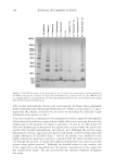
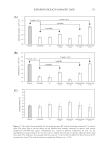
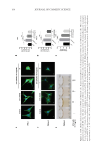
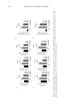
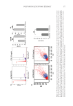
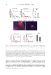
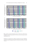
and Rab27a in PIG1 cells, while HCP treatments significantly inhibited the expression
of Rac1 and Rab27a induced by ET-1 in.a dose-dependent manner. Consistently, we also
demonstrated that HCP could inhibit the expression of Rac1 and Rab27a in MelanA cells
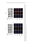
both at the mRNA and protein levels (Figures 3D–F). These findings suggest that HCP
may inhibit the transport of melanosomes in MelanA and PIG1 cells by downregulating
gene expression of Rac1 and Rab27a.
HCP INHIBITED ET-1-INDUCED ACTIVATION OF WNT/Β-CATENIN SIGNAL PATHWAY
It is reported that MITF not only controls the expression of factors related to melanin
synthesis but also plays a role in regulating the expression of Rab27A.14 Therefore, we
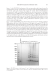
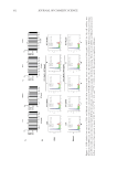
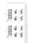
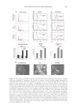
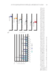
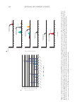
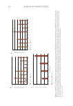
investigated the impact of HCP on the expression of MITF in melanocytes. As shown in
Figures 4A and 5A, 20 nM ET-1 notably increased the expression of MITF in PIG1 cells,
while HCP effectively reversed the increase of MITF expression induced by ET-1. Similar
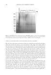
results were also observed in MelanA cells (Figures 4B and 5B). TYR, the key enzyme
involved in melanin synthesis, is recognized as a target gene of MITF. This is attributed to
the presence of an E-box motif within the TYR promoter, which facilitates the binding of
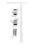
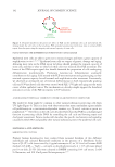
the transcription factor MITF. Consistently, the gene expression and activity of TYR were
also inhibited by HCP treatments in MelanA cells (Figure 6A–C).
Considering that the expression of MITF can be regulated by the Wnt/β-catenin signaling
pathway, we further investigated the effects of HCP on this signaling pathway. As shown
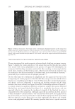
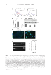
in Figures 4A and 4B, the level of phosphorylation GSK-3β and β-catenin in PIG1 cells
was significantly increased by ET-1, while 1 µg/ml and 10 µg/ml HCP treatment greatly
reversed the ET-1-induced increase of p-GSK-3β and β-catenin. GSK-3β can combine
with β-catenin to form a complex, keeping β-catenin in an inactive state. As expected,
HCP exhibited a concentration-dependent inhibitory effect on the ET-1-induced increase
in β-catenin expression. Importantly, the increase of ET-1-induced β-catenin nuclear
localization in both PIG1 and MelanA cells was greatly inhibited by HCP (Figures 4C
and 4D Figure 6C). These data indicate that HCP treatments can inhibit the activation
of Wnt/β-catenin signaling pathway induced by ET-1. Therefore, it is inferred that HCP
may suppress ET-1-induced dendrite formation in melanocytes by inactivating the Wnt/β-
catenin signaling pathway.
DISCUSSION
The metabolism of melanin is a systematic process involved in melanin production,
transport, uptake, and degradation.2 Both melanogenesis and melanin transfer contribute
to the accumulation of melanin in the skin. A previous study reported that soluble pearl
extract was able to inhibit the expression of TYR in B16 melanoma cells, indicating its
potential as a skin whitening agent.10 However, the specific impact of HCP (the most
abundant protein in pearl powder) on melanin synthesis and transfer remains unclear. In
this study, we find that HCP can reduce melanocyte dendrites and melanin secretion by
antagonizing the effects of ET-1. Additionally, HCP has shown the ability to downregulate
melanosome transport-related gene expression and inhibit the activity of the Wnt/β-catenin
signal pathway. Our results demonstrate that HCP seems to inhibit melanin transfer by
regulating the Wnt/β-catenin signal pathway. It is important to note that HCP, at a
and Rab27a in PIG1 cells, while HCP treatments significantly inhibited the expression
of Rac1 and Rab27a induced by ET-1 in.a dose-dependent manner. Consistently, we also
demonstrated that HCP could inhibit the expression of Rac1 and Rab27a in MelanA cells
both at the mRNA and protein levels (Figures 3D–F). These findings suggest that HCP
may inhibit the transport of melanosomes in MelanA and PIG1 cells by downregulating
gene expression of Rac1 and Rab27a.
HCP INHIBITED ET-1-INDUCED ACTIVATION OF WNT/Β-CATENIN SIGNAL PATHWAY
It is reported that MITF not only controls the expression of factors related to melanin
synthesis but also plays a role in regulating the expression of Rab27A.14 Therefore, we
investigated the impact of HCP on the expression of MITF in melanocytes. As shown in
Figures 4A and 5A, 20 nM ET-1 notably increased the expression of MITF in PIG1 cells,
while HCP effectively reversed the increase of MITF expression induced by ET-1. Similar
results were also observed in MelanA cells (Figures 4B and 5B). TYR, the key enzyme
involved in melanin synthesis, is recognized as a target gene of MITF. This is attributed to
the presence of an E-box motif within the TYR promoter, which facilitates the binding of
the transcription factor MITF. Consistently, the gene expression and activity of TYR were
also inhibited by HCP treatments in MelanA cells (Figure 6A–C).
Considering that the expression of MITF can be regulated by the Wnt/β-catenin signaling
pathway, we further investigated the effects of HCP on this signaling pathway. As shown
in Figures 4A and 4B, the level of phosphorylation GSK-3β and β-catenin in PIG1 cells
was significantly increased by ET-1, while 1 µg/ml and 10 µg/ml HCP treatment greatly
reversed the ET-1-induced increase of p-GSK-3β and β-catenin. GSK-3β can combine
with β-catenin to form a complex, keeping β-catenin in an inactive state. As expected,
HCP exhibited a concentration-dependent inhibitory effect on the ET-1-induced increase
in β-catenin expression. Importantly, the increase of ET-1-induced β-catenin nuclear
localization in both PIG1 and MelanA cells was greatly inhibited by HCP (Figures 4C
and 4D Figure 6C). These data indicate that HCP treatments can inhibit the activation
of Wnt/β-catenin signaling pathway induced by ET-1. Therefore, it is inferred that HCP
may suppress ET-1-induced dendrite formation in melanocytes by inactivating the Wnt/β-
catenin signaling pathway.
DISCUSSION
The metabolism of melanin is a systematic process involved in melanin production,
transport, uptake, and degradation.2 Both melanogenesis and melanin transfer contribute
to the accumulation of melanin in the skin. A previous study reported that soluble pearl
extract was able to inhibit the expression of TYR in B16 melanoma cells, indicating its
potential as a skin whitening agent.10 However, the specific impact of HCP (the most
abundant protein in pearl powder) on melanin synthesis and transfer remains unclear. In
this study, we find that HCP can reduce melanocyte dendrites and melanin secretion by
antagonizing the effects of ET-1. Additionally, HCP has shown the ability to downregulate
melanosome transport-related gene expression and inhibit the activity of the Wnt/β-catenin
signal pathway. Our results demonstrate that HCP seems to inhibit melanin transfer by
regulating the Wnt/β-catenin signal pathway. It is important to note that HCP, at a