175 POLYPODIUM LEUCOTOMOS EXTRACT
RESULTS
PLE SLOWS DOWN GROWTH AND PROTECTS THE AMPLIFYING CAPACITY OF HUMAN
EPIDERMAL KERATINOCYTES
We cultured freshly isolated keratinocytes from human epidermis in Rheinwald
conditions.25 In these conditions, cells proliferate, stratify, and differentiate. To investigate
the effect of PLE on the clonal growth capacity of epidermal stem cells, we performed
clonogenicity assays with human isolated primary keratinocytes of the skin. These assays
at low density allow monitoring the growth of epidermal stem cells since they are the
only cells capable to give rise to large proliferative colonies.26Proliferative progenitors
are committed to differentiate after a small number of divisions and give rise to small
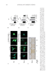
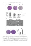
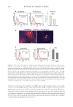
differentiated colonies. Primary keratinocytes cultured in the presence of PLE for 8 days
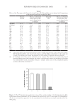
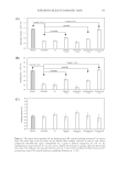
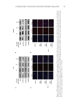
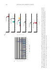
(0.8 or 1.6 mg/ml) displayed a significantly greater reduction in the size of the colonies
than controls (Figure 1A). When we examined the cultures after incubation with PLE for
8 days, we observed that cells were slightly larger and less densely packed than controls
(Figure 1B). The smaller size of the colonies upon PLE might be caused by a toxic effect or
by retarded growth. To answer this question, we plated cells for clonogenicity assays after
treatment, in the absence of PLE, to test for the later effects on the capacity of stem cells.
Interestingly, PLE-treated keratinocytes displayed a significant increase in the number of
colonies (Figure 1C).
PLE DELAYS PROGRESSION OF KERATINOCYTES THROUGH THE G2 PHASE OF THE CELL CYCLE
AND INDUCES DNA REPAIR SIGNALS ΓH2AX AND P53 INDEPENDENTLY OF DNA DAMAGE
The growth inhibitory effect of PLE suggested that it was acting on the keratinocyte
cycle. Therefore, we examined the effect on the cell cycle. As before, we treated human
keratinocytes with PLE (0.8 mg/ml) for 8 days and determined their DNA content by
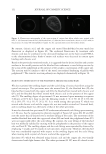
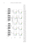
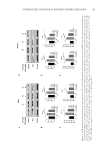
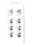
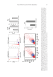
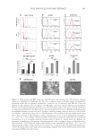
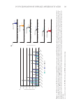
flow cytometry. We observed a striking change in the cell cycle upon PLE. G1 was
significantly diminished and G2/M was augmented (Figure 2A). We found a similar
accumulation of cells in G2/M after various periods of treatment with PLE, from 5 to 9
days (not shown). Considering that cells proliferated more slowly, this would not indicate
a cell cycle activation but rather, a delay of progression through the G2/M transition.
We observed no signs of apoptotic sub-G1 cells in the DNA content analyses, further
indicating that the growth inhibitory effect of PLE was due to a slower cell cycle. An
increase in forward scatter and side scatter was also observed within the proliferative
region, consistent with the morphology displayed in Figure 1 and with the typical cell
size increase in G2/M due to the cellular growth that takes place prior to cell division
(Figure 2B).20,26
Since keratinocytes were more clonogenic after the PLE treatment, we investigated the
expression of γH2AX by flow cytometry, a central signal in the induction of the DNA repair
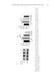
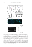
cascade.27 Interestingly, upon a similar PLE treatment as before, we observed a significant
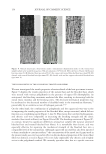
increase in the phosphorylation of H2AX (γH2AX Figure 3A and B). Since γH2AX
induces the G2/M checkpoint to allow DNA repair, in part via the p53, we analyzed the
expression of this transcription factor by Western blotting and immunofluorescence (Figure
3C and D). PLE treatment significantly induced the expression of p53 to levels comparable
to those induced by treatment with the genotoxic drug Doxorubicin (Figure 3C).
RESULTS
PLE SLOWS DOWN GROWTH AND PROTECTS THE AMPLIFYING CAPACITY OF HUMAN
EPIDERMAL KERATINOCYTES
We cultured freshly isolated keratinocytes from human epidermis in Rheinwald
conditions.25 In these conditions, cells proliferate, stratify, and differentiate. To investigate
the effect of PLE on the clonal growth capacity of epidermal stem cells, we performed
clonogenicity assays with human isolated primary keratinocytes of the skin. These assays
at low density allow monitoring the growth of epidermal stem cells since they are the
only cells capable to give rise to large proliferative colonies.26Proliferative progenitors
are committed to differentiate after a small number of divisions and give rise to small
differentiated colonies. Primary keratinocytes cultured in the presence of PLE for 8 days
(0.8 or 1.6 mg/ml) displayed a significantly greater reduction in the size of the colonies
than controls (Figure 1A). When we examined the cultures after incubation with PLE for
8 days, we observed that cells were slightly larger and less densely packed than controls
(Figure 1B). The smaller size of the colonies upon PLE might be caused by a toxic effect or
by retarded growth. To answer this question, we plated cells for clonogenicity assays after
treatment, in the absence of PLE, to test for the later effects on the capacity of stem cells.
Interestingly, PLE-treated keratinocytes displayed a significant increase in the number of
colonies (Figure 1C).
PLE DELAYS PROGRESSION OF KERATINOCYTES THROUGH THE G2 PHASE OF THE CELL CYCLE
AND INDUCES DNA REPAIR SIGNALS ΓH2AX AND P53 INDEPENDENTLY OF DNA DAMAGE
The growth inhibitory effect of PLE suggested that it was acting on the keratinocyte
cycle. Therefore, we examined the effect on the cell cycle. As before, we treated human
keratinocytes with PLE (0.8 mg/ml) for 8 days and determined their DNA content by
flow cytometry. We observed a striking change in the cell cycle upon PLE. G1 was
significantly diminished and G2/M was augmented (Figure 2A). We found a similar
accumulation of cells in G2/M after various periods of treatment with PLE, from 5 to 9
days (not shown). Considering that cells proliferated more slowly, this would not indicate
a cell cycle activation but rather, a delay of progression through the G2/M transition.
We observed no signs of apoptotic sub-G1 cells in the DNA content analyses, further
indicating that the growth inhibitory effect of PLE was due to a slower cell cycle. An
increase in forward scatter and side scatter was also observed within the proliferative
region, consistent with the morphology displayed in Figure 1 and with the typical cell
size increase in G2/M due to the cellular growth that takes place prior to cell division
(Figure 2B).20,26
Since keratinocytes were more clonogenic after the PLE treatment, we investigated the
expression of γH2AX by flow cytometry, a central signal in the induction of the DNA repair
cascade.27 Interestingly, upon a similar PLE treatment as before, we observed a significant
increase in the phosphorylation of H2AX (γH2AX Figure 3A and B). Since γH2AX
induces the G2/M checkpoint to allow DNA repair, in part via the p53, we analyzed the
expression of this transcription factor by Western blotting and immunofluorescence (Figure
3C and D). PLE treatment significantly induced the expression of p53 to levels comparable
to those induced by treatment with the genotoxic drug Doxorubicin (Figure 3C).