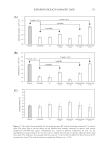
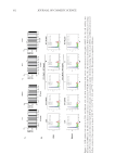
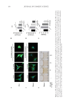
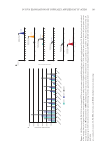
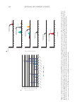
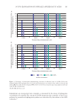
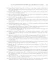
Figure
1.
HCP
exhibits
low
toxicity
to
PIG1
and
MelanA
cells.
(A)
After
incubation
with
various
concentrations
(0-100
µg/ml)
of
HCP
for
24h,
cell
viability
was
determined
by
MTS
assay.
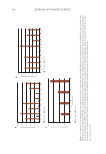
(B)
After
incubation
with
various
concentrations
(0-90nM)
of
ET-1
for
24h,
cell
viability
was
determined
by
MTS
assay.
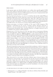
(C)
After
pre-treating
with
ET1
for
24h,
cells
were
then
treated
with
HCP
for
the
indicated
concentration
for
another
24h
before
harvesting.
Then
cell
cycle
of
PIG1
and
MelanA
were
detected
by
flow
cytometry.
Data
represent
as
mean
±
SD,
*P
0.05,
**P
0.01,
and
***P
0.001
vs
control.
Ns
represents
no
significate
difference.
162 JOURNAL OF COSMETIC SCIENCE
1.
HCP
exhibits
low
toxicity
to
PIG1
and
MelanA
cells.
(A)
After
incubation
with
various
concentrations
(0-100
µg/ml)
of
HCP
for
24h,
cell
viability
was
determined
by
MTS
assay.
(B)
After
incubation
with
various
concentrations
(0-90nM)
of
ET-1
for
24h,
cell
viability
was
determined
by
MTS
assay.
(C)
After
pre-treating
with
ET1
for
24h,
cells
were
then
treated
with
HCP
for
the
indicated
concentration
for
another
24h
before
harvesting.
Then
cell
cycle
of
PIG1
and
MelanA
were
detected
by
flow
cytometry.
Data
represent
as
mean
±
SD,
*P
0.05,
**P
0.01,
and
***P
0.001
vs
control.
Ns
represents
no
significate
difference.
162 JOURNAL OF COSMETIC SCIENCE