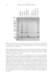
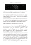
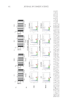
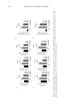
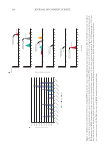
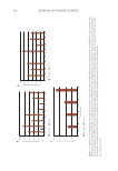
Figure
2.
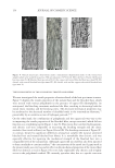
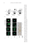
HCR
inhibits
dendrite
formation
of
PIG1
and
MelanA
cells.
Representativephotoes
of
cell
morphology
of
PIG1
(A)
and
MelanA
(C)
cells.
PIG1
cells
were
pretreated
with
20
nM
ET-1
for
24h,
then
treated
with
different
concentrations
of
HCP
for
24h.
Cytoskeleton
was
visualized
by
cell
immunofluorescent
staining
for
F-actin.
Scale
bars =
20
µ
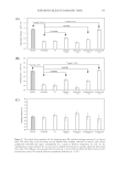
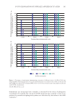
The
number
of
dendrites
were
counted
in
PIG1
(B)
and
MelanA
(D)
cells.
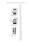
(E)
Melanin
secretion
to
the
medium
in
MelanA
cells
treated
with
20
nM
ET-1
in.the
presence
or
absence
of
HCP.
(F)
Quantitative
analysis
of
melanin
content
in
the
medium.
Data
were
expressed
mean
±
SD
from
3
independent
experiments
(n
=
3).
*P
0.05,
**P
0.01,
ET-1
vs
control
#P
0.05,
##P
0.01,
HCP
versus
ET-1.
164 JOURNAL OF COSMETIC SCIENCE
2.
HCR
inhibits
dendrite
formation
of
PIG1
and
MelanA
cells.
Representativephotoes
of
cell
morphology
of
PIG1
(A)
and
MelanA
(C)
cells.
PIG1
cells
were
pretreated
with
20
nM
ET-1
for
24h,
then
treated
with
different
concentrations
of
HCP
for
24h.
Cytoskeleton
was
visualized
by
cell
immunofluorescent
staining
for
F-actin.
Scale
bars =
20
µ
The
number
of
dendrites
were
counted
in
PIG1
(B)
and
MelanA
(D)
cells.
(E)
Melanin
secretion
to
the
medium
in
MelanA
cells
treated
with
20
nM
ET-1
in.the
presence
or
absence
of
HCP.
(F)
Quantitative
analysis
of
melanin
content
in
the
medium.
Data
were
expressed
mean
±
SD
from
3
independent
experiments
(n
=
3).
*P
0.05,
**P
0.01,
ET-1
vs
control
#P
0.05,
##P
0.01,
HCP
versus
ET-1.
164 JOURNAL OF COSMETIC SCIENCE