179 POLYPODIUM LEUCOTOMOS EXTRACT
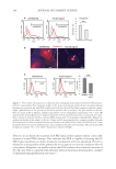
Note that γH2AX and p53 are central cellular signals responding to DNA damage to
induce DNA repair. Treatment of PLE was not expected to induce DNA damage given its
cellular protective effects shown above and elsewhere. An interesting alternative was that
PLE induces the signals independently of the damage, therefore boosting DNA repair. To
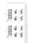
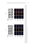
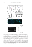
test this possibility, we performed comet assays to analyze actual nuclear chromosomal
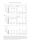
fragmentation. As shown in Figure 3E and F, the same doses of PLE that induced γH2AX
(0.8 mg/ml) did not cause an increase in DNA fragmentation. This indicates that the
significant boost of the DNA repair signals was independent of DNA damage.
PLE ATTENUATES THE DIFFERENTIATION RESPONSE INDUCED BY REPLICATION STRESS OR
SUBLETHAL UV IRRADIATION
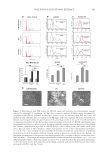
Since in keratinocytes replication stress via DNA damage triggers terminal differentiation
during proliferation, we analyzed the expression of epidermoid differentiation markers
involucrin and keratin K16.28–30 Involucrin is a precursor of the cornified envelope, and
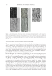
K16 is expressed in the skin during hyperplasia or other stress conditions.31 PLE reduced
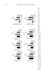
the proportion of cells expressing involucrin and keratin K16 as analyzed by flow cytometry
and immunofluorescence (Figure 4A–C).
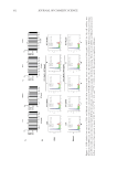
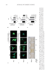
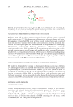
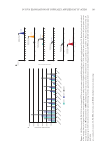
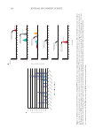
To evaluate the protective effect of PLE on skin keratinocytes on the face to UV light,
we pretreated primary keratinocytes with PLE for 6 days and then irradiated them.
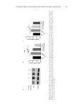
Pretreatment with PLE had only a small effect on the cell cycle profile of irradiated cells
(Figure 5A). Interestingly, the cell cycle profile of irradiated cells was very similar to the
profile induced by PLE in nonirradiated cells (compare with Figure 2A). PLE however,
induced moderately further the levels of γH2AX in irradiated cells (Figure 5B). As
mentioned above, keratinocytes respond to unrepaired DNA damage by initiating terminal
differentiation (unless exposed to acute doses of UV irradiation, which induces apoptosis).
Consistently, sublethal UV irradiation induces keratinocyte differentiation.21 To note,
treatment with PLE diminished the proportion of UV-induced terminally differentiating
cells, according to involucrin expression and cellular morphology (Figure 5C and D).
DISCUSSION
PLE REINFORCES CELL CYCLE CONTROLS
We have shown cumulative evidence indicating that PLE protects epidermal cells from
proliferative or UV-induced DNA damage by slowing down the keratinocyte cell cycle at
the G2/M transition. G1 and G2 are the major points of the cell cycle where DNA repair
is achieved.22,32 Moreover, in keratinocytes, the G2/M checkpoints control cell fate whether
to divide, to differentiate, or to undergo apoptosis.20,28 Unrepaired cells that enter mitosis
prematurely will differentiate. The cell cycle speeds up due to proliferative stimuli such
as the in vitro conditions that we have implemented in this study or upon stimuli such as
UV light.21,28 Stem cells, when activated, give rise to rapidly proliferative progeny. These
cells are already committed to differentiate terminally due to the loss of cell cycle control,
replication stress, and DNA damage.
The γH2AX signal and the transcription factor p53 both trigger the G2 checkpoints and
halt cells in G2 to promote DNA repair.22,27,33 These signals respond to DNA damage.
Note that γH2AX and p53 are central cellular signals responding to DNA damage to
induce DNA repair. Treatment of PLE was not expected to induce DNA damage given its
cellular protective effects shown above and elsewhere. An interesting alternative was that
PLE induces the signals independently of the damage, therefore boosting DNA repair. To
test this possibility, we performed comet assays to analyze actual nuclear chromosomal
fragmentation. As shown in Figure 3E and F, the same doses of PLE that induced γH2AX
(0.8 mg/ml) did not cause an increase in DNA fragmentation. This indicates that the
significant boost of the DNA repair signals was independent of DNA damage.
PLE ATTENUATES THE DIFFERENTIATION RESPONSE INDUCED BY REPLICATION STRESS OR
SUBLETHAL UV IRRADIATION
Since in keratinocytes replication stress via DNA damage triggers terminal differentiation
during proliferation, we analyzed the expression of epidermoid differentiation markers
involucrin and keratin K16.28–30 Involucrin is a precursor of the cornified envelope, and
K16 is expressed in the skin during hyperplasia or other stress conditions.31 PLE reduced
the proportion of cells expressing involucrin and keratin K16 as analyzed by flow cytometry
and immunofluorescence (Figure 4A–C).
To evaluate the protective effect of PLE on skin keratinocytes on the face to UV light,
we pretreated primary keratinocytes with PLE for 6 days and then irradiated them.
Pretreatment with PLE had only a small effect on the cell cycle profile of irradiated cells
(Figure 5A). Interestingly, the cell cycle profile of irradiated cells was very similar to the
profile induced by PLE in nonirradiated cells (compare with Figure 2A). PLE however,
induced moderately further the levels of γH2AX in irradiated cells (Figure 5B). As
mentioned above, keratinocytes respond to unrepaired DNA damage by initiating terminal
differentiation (unless exposed to acute doses of UV irradiation, which induces apoptosis).
Consistently, sublethal UV irradiation induces keratinocyte differentiation.21 To note,
treatment with PLE diminished the proportion of UV-induced terminally differentiating
cells, according to involucrin expression and cellular morphology (Figure 5C and D).
DISCUSSION
PLE REINFORCES CELL CYCLE CONTROLS
We have shown cumulative evidence indicating that PLE protects epidermal cells from
proliferative or UV-induced DNA damage by slowing down the keratinocyte cell cycle at
the G2/M transition. G1 and G2 are the major points of the cell cycle where DNA repair
is achieved.22,32 Moreover, in keratinocytes, the G2/M checkpoints control cell fate whether
to divide, to differentiate, or to undergo apoptosis.20,28 Unrepaired cells that enter mitosis
prematurely will differentiate. The cell cycle speeds up due to proliferative stimuli such
as the in vitro conditions that we have implemented in this study or upon stimuli such as
UV light.21,28 Stem cells, when activated, give rise to rapidly proliferative progeny. These
cells are already committed to differentiate terminally due to the loss of cell cycle control,
replication stress, and DNA damage.
The γH2AX signal and the transcription factor p53 both trigger the G2 checkpoints and
halt cells in G2 to promote DNA repair.22,27,33 These signals respond to DNA damage.