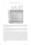
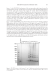
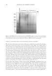
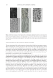
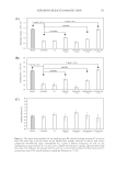
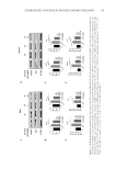
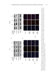
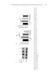
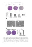
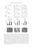
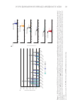
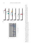
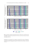
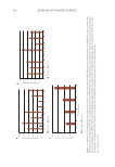
Figure
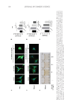
4.
HCP
inhibits
the
activity
of
Wnt/β-catenin
pathway.
PIG1(A)
and
MelanA
(B)
cells
were
pretreated
with
20
nM
ET-1
for
24h,
then
treated
with
different
concentrations
of
HCP
for
24h.
β
p-GSK-3βand
MITF
protein
expression
were
determined
by
Western
blot
analysis.
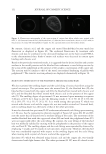
HCP
treatment
leads
to
a
reduction
in
the
nuclear
localization
of
β
Immunostainings
of
β
of
PIG1
and
MelanA
cells
after
20
nM
ET-1
treatments
with
or
without
HCP.
Nuclei
were
stained
with
4’,6-diamidino-2-phenylindole
(DAPI)
blue.
Scale
bars =
100
µm.
167 Hydrolyzed Conchiolin Protein Inhibits Melanin
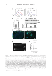
4.
HCP
inhibits
the
activity
of
Wnt/β-catenin
pathway.
PIG1(A)
and
MelanA
(B)
cells
were
pretreated
with
20
nM
ET-1
for
24h,
then
treated
with
different
concentrations
of
HCP
for
24h.
β
p-GSK-3βand
MITF
protein
expression
were
determined
by
Western
blot
analysis.
HCP
treatment
leads
to
a
reduction
in
the
nuclear
localization
of
β
Immunostainings
of
β
of
PIG1
and
MelanA
cells
after
20
nM
ET-1
treatments
with
or
without
HCP.
Nuclei
were
stained
with
4’,6-diamidino-2-phenylindole
(DAPI)
blue.
Scale
bars =
100
µm.
167 Hydrolyzed Conchiolin Protein Inhibits Melanin