94 JOURNAL OF THE SOCIETY OF COSMETIC CHEMISTS jects and on the shaved skin of several laboratory animals, result- ing in typical dandruff scales. In the human tests the chest area was used and also dandruff-free scalps. Using whole cultures of P. ova/e originally isolated from infectious dandruff, the infection was repro- duced in 100 per cent of the scalps inoculated and 79 per cent of the chest areas. Typicaldandruff scales resulted from these inoculations and P. ova/e was observed in these scales by microscopic examination. As a final step, P. ovale was then reisolated from the dandruff scales produced by these artificial in- oculations. For the first time all of the requirements of Koch's pos- tulates had been fulfilled to prove the exact cause of this disease. It is of interest to note that they were able to reproduce the disease by inoculating P. ovale into the skin of animals, especially rabbits and guinea pigs, and to reisolate the organism from the typical branny scales. Control areas similarly treated but without inoculation with P. ova/e did not show dandruff scales. Also they were unable to reproduce the disease by the use of common yeast and another micro- organism isolated from the scalp which was obviously a saprophytic contaminant and not infectious. Histopathologic examinations were made of biopsies of the skin of animals infected with P. ova/e and specific characteristic reactions were noted. From these observations it was noted that "... the induced dermatitis was the result of the invasion of the epidermis and the hair follicles by these organisms." This important discovery by Moore and his associates has been confirmed independently by Durfee and Cousins (21). These investi- gators, using the methods developed by Moore, isolated pure cultures of Pityrosporum ova/e from dandruff scales, reproduced the infectious condition by inoculating pure cul- tures of this organism into the scari- fled skin of rabbits, and then re- isolated the organism from scales formed on the infected rabbit skin. Control areas on these same rabbits which were not inoculated with Pityrosporum ova/e showed no in- fection, no s•aling, no trace of dandruff. This work offers addi- tional confirmation of the previous results announced by Moore and Kile and their associates and offers further evidence that Pityrosporum ova/e is a causative agency of infec- tious dandruff. The morphological characteristics of this microiSrganism were origi- nally described by Unna as follows: cells short cylindric, 1 X 2ta, either swelling to spherical cells, 2-3 times normal, or becoming flask-shaped, rarely pyriform or the shape of a dumbbell, or even elongating to 2-3 times normal length, 1.5-3 X 2-4u. This description is still recognized by mycologists as characteristic of P. ovale. The detailed morphological and cultural characteristic as described by Moore (1935) are as follows: "On Sabouraud agar, colony flat,

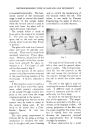
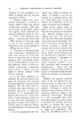
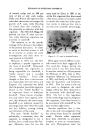
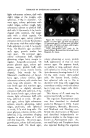
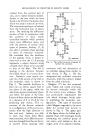
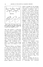
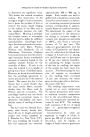
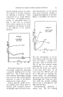
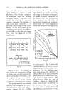
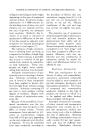
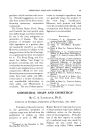
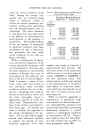
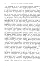
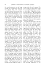
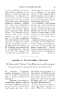
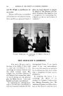
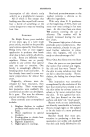
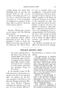
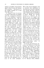
ETIOLOGY OF INFECTIOUS DANDRUFF 95 light ochraceous salmon, dull with slight ridges at the margin, cells spherical, 3-10• in diameter. On wort agar, colony pulvinate with radial ridges, surface rough, light ochraceous salmon to pinkish buff, dull cells 3-15u mostly 4-5u, bottle- shaped cells common, the larger cells with a thick capsule. On malt extract agar, colony pinkish cinnamon with a circular flat plateau in the center with fine radial ridges. Cells spherical or ovid, 2-7u mostly 4-5u. On Raulin's agar and Rich- ard's agar, growth similar but very poor. On cornmeal agar, colony ochraceous buff, slightly glistening, ridges faint, margin ir- regular. Long cells occasional. On potato glucose agar, colony pul- vinate, waxy, pinkish buff, cells spherical to ovoid, mostly bottle- shaped 2-6/• mostly 4/•. On Maneval's modification of Gorod- kova agar, colony similar, light ochraceous salmon, cells similar but stone long cells present. On lac- tose broth agar and nutrient agar, colony flat, or s}ightly elewtted, cinnamon buff, cells spherical, 2-5/•. On lactose broth agar, col(my pul- vinate, light ochraceous buff, cells 4.5•, spherical or ovoid. On glyc- erol agar, colony light ochraceous salmon, smooth, glistening, radial ridges, pulvinate, cells spherical or ovoid, 2-7u with long cells. On yeast glucose, colonies similar, cells spherical, 2-5u, sometimes clinging together in short chains of 3 cells with few long cells. On serum agar, growth poor, colony dull and pasty, ochraceous buff. On blood agar, Figure III.- Colony cultures on different media: (1) wort agar, (2) malt extract agar, (3) cornmeal agar, I. to R top row, (4) Sabou- raud's agar, (5) brain veal agar, and (6) glyc- erin agar L to R bottom row. These photographs were made by Dr. Morris Moore, Barnard Free Skin and Cancer Hospital, St. Louis. colony glistening or waxy, pinkish buff, appearance of that on malt extract agar. On peptone broth, no pellicle or ring, sediment shows cells spherical to ovoid, 3 -9• mostly 5u in diameter, fi•rming chains 3-5 X 12-15u with many thick-walled cells. On lactose broth, similar, but spherical cells up to 12u in diameter with long cells suggesting catenulate conidia, ovoid cells about 6u in long axis, larger cells thick- walled." This history of P. ova/e may be summarized briefly as follows: The organism Pi(¾rosporum ova/e was first described in dandruff scales by Malassez in 1874. Later investigators, Benedek, Bizzozero, and MacLeod and Dowling, con- firmed the observations of Malassez and described independently or- ganisms similar to the fungus now known as Pityrosporum ova/e. Ac- ton and Panja and Templeton have also reported finding this organism
Purchased for the exclusive use of nofirst nolast (unknown)
From: SCC Media Library & Resource Center (library.scconline.org)