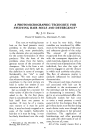
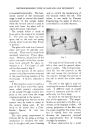
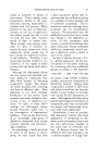
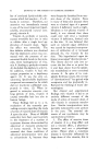
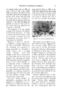
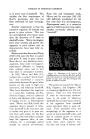
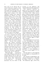
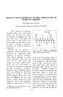
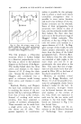
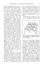
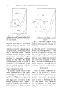
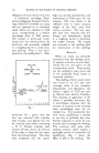
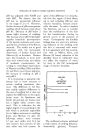
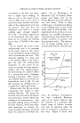
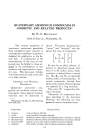
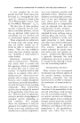
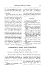
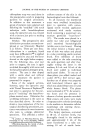
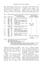
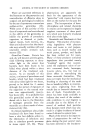
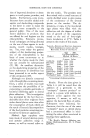
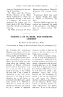
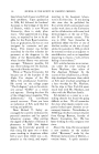
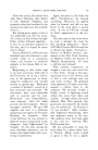
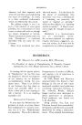
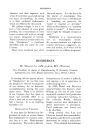
100 JOURNAL OF THE SOCIETY OF COSMETIC CHEMISTS application of paper partition chro- matography (9, 10). The amino acid whose side chain is probably most important in de- termining the properties of kera- tin is cystine, half of whose side chain is shown within the dotted box in Fig. 1. The free valence of the sulfur atom is attached to an- other sulfur in a similar side chain on an adjoining molecule. Cys- tine in this way transforms the keratin molecule into a more or less rigid three dimensional network. These interchain bridges of covalent bonds prevent the solution of hair or wool keratin in solvents which cannot chemically break such bonds, and also resist the slippage of the peptide chains pastone anotherwhen the fiber is stretched or deformed. X-RAY DIFFRACTION EVIDENCE OF KERATIN STRUCTURE When groups of molecules are arranged so that there are regularly Fig. 2.--X-ray fiber diagram of human hair. recurring distances between cer- tain atoms, the material which they compose can diffract x-rays in such a way that the x-ray beam is strongly reflected in specific direc- tions. A photographic record of these strong reflections is called a diffraction pattern. From the loca- tion and intensity of the reflections in such a pattern, the interatomic spacings in the irradiated material can be determined.* For many crystalline compounds, where the regularity of arrangement is excep- tionally high, the molecular struc- ture may be calculated directly from x-ray data independently of chemical evidence, but information from hundreds or thousands of diffraction spots must be obtained and used. Ordinary keratin of hair or wool gives the type of diffraction pattern shown in Fig. 2. Instead of the numerous, small, well-defined spots which are found in the diffraction patterns of crystalline materials of low molecular weight, this pat- tern has only two symmetrical pairs of reflections. The fact that these spots are diffuse and. are spread out into arcs shows that the regu- larity of the molecular arrangement which produced them is not very high. The information which can be obtained directly from this dia- gram is that there is a repeat dis- tance along the fiber axis of 5.1 •, or a multiple of this distance, cal- * A good introduction or review for this subject is a book by K. Lonsdale, "Crystals and X-rays," New York, Van Nostrand (1949).

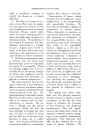
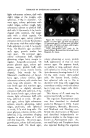
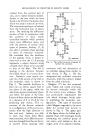
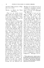
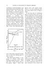
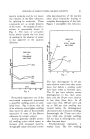
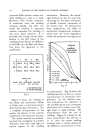
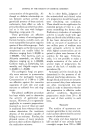
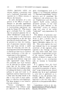
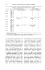
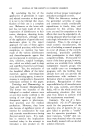
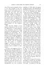
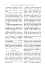
DEVELOPMENTS IN STRUCTURE OF KERATIN FIBERS 101 culated from the vertical pair of arcs, and a repeat distance perpen- dicular to the axis which we have found to be 9.0 • for the human hair which was used in some of our work. The transverse spacing is calculated from the horizontal pair of heavy spots. By studying the diffraction pattern of hair in comparison with the patterns of more nearly crystalline keratins which produce many more diffraction spots, and with the patterns of certain other types of proteins, Astbury (3, 4) was able to interpret these spacings in terms of molecular structure. He concluded that the backbone of the peptide chain is folded in some way so that the 5.1 • spacing represents a repeat distance along a folded chain whose axis is parallel to that of the fiber. The 9.0 • spacing is the distance between the folded chains in a lateral direc- tion. Astbury's most recent con- cept (2) of the nature of the fold in the molecular chain is shown in Fig. 3. The peptide backbone is bent into an almost square fold in the plane of the paper, while the side chains project alternately above and below the plane of the folds. Layers of these chains are held apart at a distance of 9 to 10 • by the projecting side chains. The oc- currence of the keratin molecule in a configuration of this sort is con- sistent with the high elasticity of hair and wool. Astbury was able to show that when these fibers were stretched, their diffraction pat- tern was changed from the one shown to another whose spacings were up uP [] = SIDlECHAIN Fig. &--The folded structure proposed by Astbury for a-keratin. consistent with the dimensions of an unfolded zigzag chain similar to that shown in Fig. 1. He has designated this unfolded structure fi-keratin, while the ordinary folded form he called c•-keratin. Huggins (17) has also proposed some folded and coiled structures for keratin molecules which will fit the scanty x-ray evidence. He has particularly emphasized the role which hydrogen bonds play in stabilizing the molecular con- figuration. The type of structure which Huggins regarded to be most likely for normal hair keratin is shown in Fig. 4. The upper part of the figure shows the folded poly- peptide chain with hydrogen bonds connecting the carbonyl group of one amino acid residue to the ni- trogen atom of an adjacent residue.
Purchased for the exclusive use of nofirst nolast (unknown)
From: SCC Media Library & Resource Center (library.scconline.org)