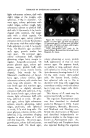
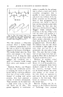
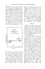
104 JOURNAL OF THE SOCIETY OF COSMETIC CHEMISTS secondary folded structure of Hug- gins is not correct. DEGREE Or ORDER AND FIBER HISTOLOGY There are two further factors which make the structure of hair fibers even more complicated than it may have seemed above. These factors are the degree of order and the histology of the fiber. In the first part of this paper, only those regions of the fiber were being considered which are suffi- ciently well-ordered to scatter x-rays coherently. However, molecules formed and arranged as regularly as the models discussed above do not make up the whole of a keratin fiber. There are parts of the fiber where the coiled molecules may be twiited or tangled, or where the coils themselves are not perfectly formed. There is no actual bound- ary between these so-called amor- phous portions of the fiber and those which are more perfectly arranged. The arrangement of the chain mole- cules varies gradually from very good order to almost complete disorder. The boundary of ordered regions would depend upon the method used to look for it, just as do measurements of atomic radii or the"crystallinity"ofcellulose. Un- like the case of cellulose, however, no satisfactory method of any sort has been found for getting a rea- sonable measure of the amount of ordered material in keratin. An estimate of 25 per cent "crystal- linity" for wool has been made by Consden (8), and Hailwood and Horrobin (12) calculate the ratio of crystalline to amorphous material to be about 0.76-0.92, but it is hard to say just how meaningful these figures are. The histology of wool and hair fibers is perhaps a little better understood. The center of the fiber contains spindle cells. a few microns thick and about 100 t• long packed closely together. The out- side of the fiber is sheathed in a layer of flat scale cells Which overlap each other to a degree which de- pends on the type of fiber. Be- tween these two portions of the fiber, and perhaps among the central cells, occurs a material which has been referred to as the "cementing substance." The layer of this sub- stance lying between the scale cells and the center cortical cells may be isolated in the form of a continuous tubule (18, 11, 1), known as the subcuticle, and has a chemical behavior very different from that of the scales and cortex.* Although the amino acid distribution in this mem- brane may not be the same as that of the whole fiber (1), it does not differ enough to account for the observed chemical difference. Hap- pey (15) has found by x-ray examina- tion that the tubules contain chain molecules which are not coiled, but are in a straightened, or /Lkeratin configuration. Although in his ex- * Note added in proof: Recent work in this laboratory by Dr. E. H. Mercer indicates that the subcuticle consists of resistant cor- tical c.ells, so that the name "subcuticle" is a misnomer. The epicuticle of the fiber (20, 21) is usually also present in chemical preparations of the subcuticle.

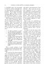
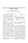
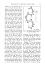
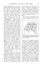
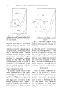
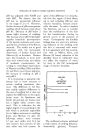
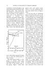
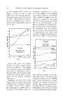
DEVELOPMENTS IN STRUCTURE OF KERATIN FIBERS 105 periment the structure may have been changed in the isolation of the membrane (25), it is likely that some difference in molecular ar- rangement accounts for the chemi- cal behavior of this portion of the fiber. Long before the subcuticle had been isolated and studied, it had been found (29) that the cortical cells had x-ray diffraction and mechanical properties comparable to those of the whole fiber, while the scale cells consisted of much more disordered material. The kind of order in the molecular chains and its extent certainly varies among scale cells, cortical cells, and sub- cuticle, and at present we have no quantitative measure of the dis- tribution of ordered material among these components. EFFECT OF LiBr os KERATIN F•BERS Differences in chemical behavior can be observed between the re- gions of the fiber having high and low order. even though these regions cannot be located histologically. One of these differences is in the reaction of the fiber to the applica- tion of LiBr solutions of varying concentration (13). If a hair fiber is immersed first in water and then stepwise in aqueous LiBr solutions of gradually increasing concentra- tion, the normal x-ray diffraction pat'tern rather suddenly disappears when the salt concentration reaches about 5 M. Swelling measure- ments (28) have shown that the rate of volume change with con- centration is larger between 5 M and 9 M than above or below these concentrations. The change in x- ray diagram means that, at more dilute concentrations, LiBr solu- tions are able to penetrate only those portions of the fiber which are so disordered that they do not contribute to the x-ray diffraction pattern. At 5 M concentration and above, however, the solution can enter the "crystalline" regions of the fiber and spread the chains apart in such a way that their arrangement is no longer sufficiently regular to diffract x-rays. This disarrangement probably does not disturb the coiling of the mole- cules. At high concentrations, where the swelling levels off, the distension of the fiber is reaching the limit imposed by the co-valent cystine cross-links between chains. This whole process is reversible. If the fiber is moved into less and less concentrated solutions, the swell- ing decreases and the x-ray pattern reappears. Both phenomena show hysteresis effects. The normal x- ray diffraction pattern reappears at a lower concentration than that required to alter it originally, and the fiber does not quite contract to its original volume. This change in the structure of keratin fibers manifests itself in their physical properties. If the force to extend a hair or wool fiber 20 per cent is measured as the fiber is moved through a series of LiBr solutions, the results shown in Fig. 6 are obtained. As the fiber is swollen at low concentrations by
Purchased for the exclusive use of nofirst nolast (unknown)
From: SCC Media Library & Resource Center (library.scconline.org)