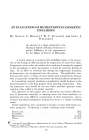
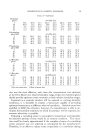
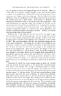
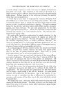
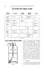
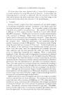
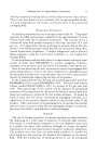
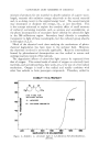
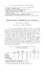
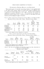
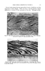
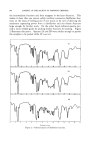
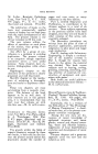
PERCUTANEOUS ABSORPTION OF VITAMIN A 371 Histological studies were made by Profi Jerome P. Parnell (State Univer- sity of New York College of Medicine at New York City). Histological changes of the skin of test rats are pictured in Figs. 1-9. Figure 3.--4Dne week after treatment, untreated site. Rat 63 days old. Note the very dense connective tissue and complete loss of dermal fat that characterizes a hypovitaminotic A skin condition (X140). Figure 4.--One week after treatment, site of application. Rat 63 days old. Note some thickening of the epidermis (X 140).

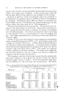
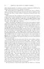
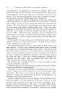
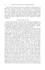
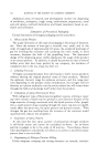
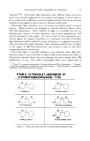
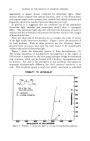
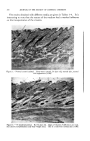
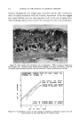
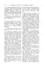
372 JOURNAL OF THE SOCIETY OF COSMETIC CHEMISTS ß . .. ...• •.,• •,, ... .5.k•. • •-•. • Figure 5.•Three weeks after treatment, untreated site. Rat 77 days old. The epidermis is capable of supporting few cells. The cells lose their nutritional support, become keratinized and slough (X 100). Figure 6.--Three weeks after treatment, site of application. Rat 77 days old. The epi- thelium is thick, consisting of many layers of large cells. Mitotic figures are abundant in the stratum germinativum, and the hair bud is beginning its downward growth (X 100).
Purchased for the exclusive use of nofirst nolast (unknown)
From: SCC Media Library & Resource Center (library.scconline.org)