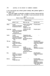
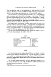
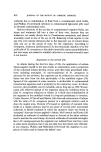
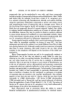
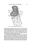
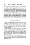
434 JOURNAL OF THE SOCIETY OF COSMETIC CHEMISTS a protofibrillar substructure has been obtained by several investigators (42) who succeeded in isolating fine filaments, about 2 nm in diameter, from e-keratin preparations fragmented by ultrasonic irradiation. However, it is contended that the filaments observed in these preparations almost cer- tainly result from cellulosic contaminants (41, 43). Moreover, experiments by Sikorski and associates (44) on the interpretation of high magnification electron micrographs of keratin fibre sections indicate that the distinctions between the microfibrillar patterns of various cortical cells may not be as simple as has been indicated. They regard the 'microfibril' as a manifestation of an average mode of aggregation in situ of protofibrils. The medulla The medulla is a histological component situated near the centre of the fibre in many keratin fibres such as human hair, but it does not occur in fine wool fibres (45). It is formed from an axial stream of cells, the contents of which shrivel up during dehydration leaving a series of vacuoles along the fibre axis. Many variations occur in the shape and size of this part of the fibre. It has been usual, however, to concentrate studies on wool fibres in which medulla is either absent or present in only small amounts. In any case, the medulla is believed to make little contribution to the chemical and mechanical properties of the fibre. That the material of the medullary cells differs from that of the surrounding cortical cells is indicated by the differences in staining characteristics of the two types of cells. Recently, amino acid analyses of the separated medullary cells from rabbit, kangaroo, and platypus hair show that they contain about 1 residue in 4-5 of glutamic acid, 1 in 9 of citrulline, 1 in 12 of leucine, 1 in 14 of glycine and only 1 in 35 of cystine (46). The citrulline is covalently bound in the peptide linkage in the proteins (47). It has been shown that the presence of N6-q, - glutamyllysine cross-link in hair and quill medulla protein of mammalian species is a general phenomenon (48). The structure of human hair An insight into the histological structure of human hair has been gained from the electron microscopic observations in this laboratory of sections of ether-degreased fibres stained with various heavy metal compounds (49). Human hair, like most other keratin fibres, consists of a central core of long spindle-shaped interdigitating keratin-filled cortical cells this core is bounded by a sheath of overlapping leaf-like cuticle cells. Perhaps the most

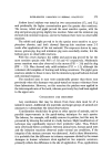
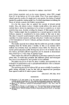
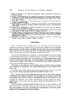
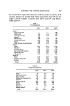
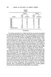
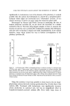
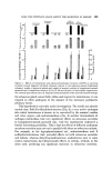
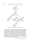
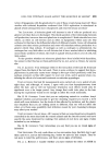
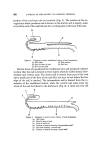
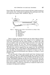
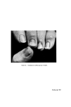
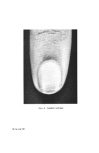
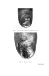
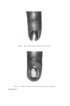
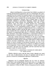
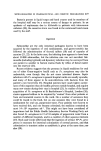
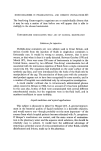
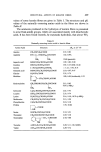
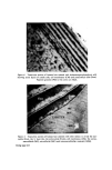
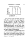
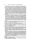
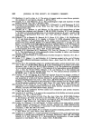
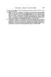
Figure 4. Transverse section of human hair stained with dodecatungstophosphoric acid showing seven layers of cuticle cells, cell membranes (CM) and paracortical cells (Para). Pigment granules (PG) in the cortex are black. Figure 5. Transverse section of human hair stained with silver nitrate to reveal the cxo- cuticle (Exo), the 'a' layer (A), the endocuticle (Endo), cell membranes (CM), the cortex, macrofibrils (MF), microfibrils (MiF) and intermacrofibrillar material (IMM). Facing page 435
Purchased for the exclusive use of nofirst nolast (unknown)
From: SCC Media Library & Resource Center (library.scconline.org)