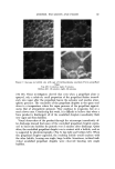
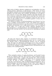
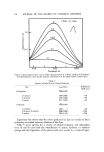
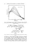
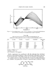
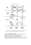
89. JOURNAL OF THE SOCIETY OF COSMETIC CHEMISTS Facial wrinkles are produced by repeated and habitual contraction of the underlying muscles of facial expression. When the facial muscles contract, the muscle shortens without a corresponding shortening of the overlying skin, thereby producing a wrinkle. The skin adapts itself by forming folds at right angles to the line of contraction of the underlying muscle (2). The supraorbi- tal wrinkle lines are caused by the contraction of the frontalis muscle which is inserted into the skin of the lower forehead. The transverse lines of the fore- head are due to the contraction of the frontalis muscle, and as the skin adapts itself to contraction of the muscle fibers, folds of excess skin are formed. In the upper eyelid, fine perpendicular strands of connective tissue terminate in the dermis to form the tarsal fold at the insertion of the levator palpebrae superioris. Similar tendinous insertions in the lower lid create the fine hori- zontal wrinkle lines which are accentuated by the contraction of the orbicu- laris oculi muscle (3). Other superficial facial muscles and their insertions are responsible for the other characteristic grooves and creases which are ap- parent in the face when it is not at rest. In later years, even when at rest, the face shows these lines and furrows quite well. Other factors, in addition to the pull of muscles, that result in the forma- tion of grooves and furrows which .are called wrinkles include the distribution and ratio of collagen and elastic fibers, the thickness of the skin and the amount of underlying fat, the water content of the skin, the activity of the appendegeal glands, and the biochemical changes of the connective tissue ground substance which occur with aging (4). Some histological studies of skin have shown that the process of aging is accompanied by a thinning of the epidermis with reduction in the size of the rete ridges. Volarelli (5) found that at the deepest portion of a wrinkle there was extreme thinning of the epidermis with only two or three layers of malpighian cells. The dermal papillae were completely absent, and the epi- dermal-dermal junction had become a straight line. On the other hand, Free- man et al. (6) did not find a statistically significant change with age in thickness of the epidermis of unexposed skin of the buttocks in individuals ranging in age from 25 to 76 years. With age, both collagen and elastic fibers undergo a change, most marked on exposed portions of the body. In exposed areas of the skin, .the fibers in the subpapillary layer show an increase in basophillic staining when hema- toxylin and eosin are used (7). The collagen fibers appear clumped and show basophilic degeneration. The elastic tissue also stains poorly, and has been described as being "fragmented." Examination of elastic ,tissue in thick sec- tions demonstrates that in areas of elastosis the fibers are increased in number and are tortuous, the latter accounting for the appearance of h'agmentation .as the microtome knife slices through the tissues (8). Unna, in 1896, described the alteration in connective tissue which is seen in aged (especially exposed) skin, and he felt that the sinfilarity of staining characteristics which devel- oped represented a fusion of collagen and elastin. Unna used the various

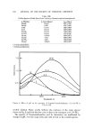
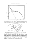
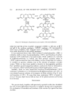
HISTOPATHOLOGY OF WRINKLES 83 descriptive terms, collacin, elacin, and collastin, •vhen discussing the altered connective tissue (9). Rather than entering into a detailed discussion of the complex and somewhat controversial changes which fake place in the corium as a function of aging, for our purpose the significant change is a loss of elasticity to the skin. The normal distensibility of the corium is due to the presence of the collagen fibers, although collagen itself is not elastic. The elastic fibers which stretch tend to prevent overextension of the collagen net- work (10). In aged skin, due to the alteration in both collagen and elastin, the normal elasticity of the skin is lost. Measurements of deformation of the skin in response to indentation sho•v that, while a decrease in normal elastic fibers correlates with the initial laxity of senile skin •vhen a deforming force is applied, as the force increases senile skin offers greater resistance than youthful skin (11). Increased intert•vining of collagen and decreased amounts of ground substance are felt to account for the reduced extensibility and in- creased tensile strength of elderly skin. Hydration is .a factor in •vrinkling, for the stratum corncure becomes de- hydrated .and brittle with aging, in large •neasure as a result of decreased wa•er-holding capacity (11). Some of the fine wrinkling seen in senile skin is attributable to this dryness of the horny layer, although the maior portion is due to the atrophy of the derreal papillae as discussed above. Also con- tributing to the decreased capacity of the skin to hold •vater is an involution of the sebaceous glands, and a reduction in the secretion of s•veat. Of the many and various changes which culminate in wrinkling, the altera- tions in ground substance appear to have especial importance to us. Such changes involve both the structural alterations of collagen and elastin and the permeability of the skin. Ma and Cowdry (12) noted a lack of clarity in the ground substance of senile skin •vhich was not apparent in the skin of younger individuals. Senile skin also shows an enhanced diffusibility of in- jected substances and a greater enhancement of spreading with the hyalu- ronidase (13). Smith et al. (14) noted a decrease in hexosamine, acid muco- polysaccharides, hyaluronic acid, and chondroitin sulfate with age. The changes in collagen which occur •vith age have been summarized by Gross (15). The fibril diameter increases with maturation but decreases slow- ly during senescense. There is an increase .of crystallinity. There is decreased swelling when collagen is subjected to dilute acids and there is a decrease in soluble collagen in acids. Resistance to collagenase is greater less base-bind- ing capacity, lower hexosamine-collagen ratio, higher hydroxproline content, and lower urinary hydroxyproline have been observed. MATERIALS AND METHODS Wrinkle specimens were obtained from 6 males and 6 females. The age range •vas 40 to 58 years. Wrinkle sites were primarily from the face, periorbi- tal region, forehead, neck, and, from one patient, the popliteal space. The
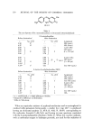
Purchased for the exclusive use of nofirst nolast (unknown)
From: SCC Media Library & Resource Center (library.scconline.org)