j. Soc. Cosmet. Chem., 30, 375-384 (December 1979) Comparison of guinea pig and fetal hog skin NANCY F. WOLEJSZA and VERA R. USDIN, Gillette Research Institute, 1413 Research Boulevard, Rockville, Maryland 20850. Received December 18, I978. Presented at the Annual Scientific Meeting, Society of Cosmetic Chemists, December 1978, New York, New York. Synopsis PROPERTIES of adult GUINEA PIG and of FETAL HOG SKIN were EVALUATED to select an appropriate model for human skin, to be used in vitro for studies of percutaneous penetration of topically applied compounds. Amino acid composition and FTIR (Fourier Transform Infra Red) spectra of isolated stratum corneum were qualitatively similar to human stratum comeurn. The major differences were in permeability, and in the relative amounts of ether and water extractable compounds in the stratum corneum. INTRODUCTION Since human skin is not readily available for in vitro studies in some laboratories, including ours, animal models must frequently be used. The suitability of a particular animal model depends on the properties studied, and its availability. The present study evaluates the permeability, amino acid composition, and content of ether- and water-extractable materials of adult guinea pig and fetal hog skin, both of which are easily obtainable in large quantity. The effects of storage were investigated by comparing the permeability of stored (-80øC) and fresh guinea pig skin. Other types of pretreatment (for example, method of hair removal) which must be standardized to obtain reproducible results were evaluated. MATERIALS SKIN SAMPLES Whole fetal hog skin (near full term) was purchased from Pel-Freez Biologicals Inc. (Rogers, Arkansas), shipped frozen and stored at -80øC until use. Whole guinea pig skin was obtained from an in-house colony of young adult male Hartley guinea pigs. The animals were sacrificed by CO2 inhalation, hair was partially removed with electric clippers, and the abdominal skin excised and either used fresh or stored at -80øC. 375

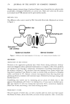
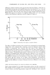
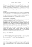
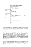
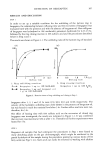
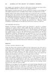
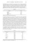
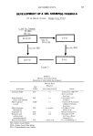
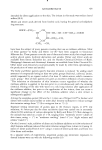
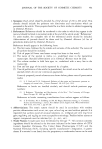
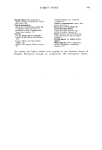
376 JOURNAL OF THE SOCIETY OF COSMETIC CHEMISTS Human stratum corneum (tops of sunburn blisters) were donated by two subjects who accidentally overexposed themselves to summer sun. These were used only for amino acid analysis and FTIR analysis not for permeability studies. DIFFUSION CELLS The diffusion cells, custom made by PGC Scientific (Rockville, Maryland), are shown in Figure 1. • Rubber cap Skin Sampling port Satura -•Ring salt solution Epidermal chamber Dermal chamber Figure 1. Diffusion cells. Inside diameter, 1.6 cm area, 2 cm2 volume of each chamber, 6 mi. METHODS PREPARATION OF SKIN SAMPLES Periderm is the uppermost epidermal layer which forms during embryonic develop- ment and disappears when epidermal keratinization is complete. Cells of the periderm are reported to go through a rudimentary development resembling keratinization of the stratum comeurn (1). The periderm layer was separated from thawed whole fetal hog skins by peeling. It was floated on water to remove folds, air-dried on a fiberglass screen, and stored at 4øC. Stratum comeurn was separated from fetal hog skins and guinea pig skins by treatment with ammonia vapor, followed by thorough rinsing with distilled water (2). Crosslinking was carried out by immersing pre-cut pieces of whole skin in 10% aqueous formaldehyde, pH 7.0 for 1 hr, followed by a 15-rain wash in distilled water. Hair was removed in vitro by treating skin specimens with a commercial depilatory (Nair ©) for 20 rain, and rinsing 2 rain under running tap water. PERMEABILITY STUDIES One-inch squares of whole skin, cut with a stainless steel die, were used. Each square was floated on water, dermis side down, to ensure similar hydradon. After 1 hr the skin
Purchased for the exclusive use of nofirst nolast (unknown)
From: SCC Media Library & Resource Center (library.scconline.org)